Fig. 6.1
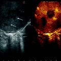
An image obtained during CEIOUS in a patient with colorectal liver metastasis. A harmonic mode image is shown in the left side and a fundamental B-mode image is shown in the right side simultaneously. A metastatic tumor in the liver is delineated more clearly in the harmonic mode image (arrows)
The vascularity of only one focal liver lesion can be examined following an injection of contrast agent. The vascularity of additional focal liver lesion can be examined as follows if this lesion appears hypoechoic during the Kupffer phase. Subsequent to the Kupffer phase scanning, Sonazoid is injected once again while the new target lesion is visualized in the ultrasound image (Fig. 6.2). This procedure enables early enhancement, if present, to be confirmed within the clearly demonstrated hypoechoic lesion. The vascularity of multiple lesions can be examined in this manner; this procedure is known as “defect reperfusion” [6]. The advantage of this technique is that the target lesion is rarely missed because it is clearly delineated as hypoechoic until the injected microbubbles reach the liver parenchyma.
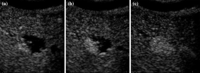
Fig. 6.2
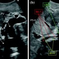
A new hypoechoic lesion was found in segment 4 during the Kupffer phase of CEIOUS in a patient with a classical HCC tumor. It was examined during the vascular phase following a repeat injection of Sonazoid. a−c Contrast enhancement appeared in the peripheral part of the lesion and gradually extended toward the center of the lesion. The lesion was occupied with contrast enhancement about 1 min after contrast injection
6.3 Hepatocellular Carcinoma
CEIOUS provides surgeons with additional information regarding new lesions found during IOUS. As described in Chap. 5, Torzilli et al. reported the usefulness of CEIOUS using SonoVue as an alternative means of differentiating HCC from benign lesions among such newly identified lesions [7]. Sonazoid, which became commercially available later than SonoVue, is also expected to be useful in a similar manner, especially with its advantage of enabling Kupffer imaging. Kupffer imaging in CEIOUS using Sonazoid is further expected to contribute to searches for new HCC tumors that were missed by IOUS and preoperative imaging studies.
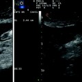
At the University of Tokyo Hospital, we conducted a prospective clinical study assessing the usefulness of CEIOUS in HCC patients [8]. A total of 192 consecutive patients were enrolled. The vascularity of one representative lesion, preferentially a new lesion found during IOUS, was examined after the intravenous injection of Sonazoid (vascular phase). Approximately 15 min after the vascular phase, total liver scanning in the harmonic mode was commenced (Kupffer phase). One additional injection of Sonazoid was allowed to examine the vascularity of another lesion, if necessary. A tentative diagnosis of HCC was made when a lesion was either hypervascular during the vascular phase or hypoechoic during the Kupffer phase (Fig. 6.3). Any lesion that was newly detected during the Kupffer phase of CEIOUS was tentatively diagnosed as HCC. A final diagnosis of HCC was made based on the results of a histological examination or dynamic computed tomography findings obtained during the 12-month postoperative period. During the study period, 79 new lesions were identified during IOUS in 50 patients (26 % of all patients), 17 of which (22 %) were finally diagnosed as HCC. The sensitivity, specificity, and accuracy of CEIOUS for differentiating HCC among the 79 IOUS-new lesions were 65, 94, and 87 %, respectively. These figures were considered satisfactory because the median diameter of the 79 lesions was 0.7 cm and all the lesions were considered so obscure that all preoperative imaging studies had missed them. Furthermore, 21 additional new hypoechoic lesions were found in 16 patients, of which 14 lesions (67 %) in 11 patients were finally diagnosed as HCC. Another advantage of the Kupffer phase of CEIOUS is that it sometimes clearly visualizes those HCC tumors which are diagnosed in preoperative images but not seen during conventional IOUS (Fig. 6.4). Surgeons cannot resect such tumors unless they can be seen in CEIOUS. Thus, CEIOUS using Sonazoid appears to enable a more accurate intraoperative staging of patients with HCC.
Get Clinical Tree app for offline access
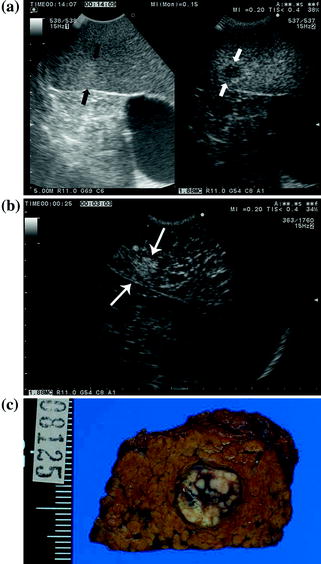
Fig. 6.3
A new focal liver lesion was found during conventional IOUS in segment 6 of the liver and was subsequently examined by CEIOUS. a In the left sided fundamental B-mode image, a hyperechoic lesion (black arrows
 Technical Tricks to Start Exploring the Liver with Ultrasound
 Diagnosis and Staging: Contrast-Enhanced Intraoperative Ultrasound (CEIOUS) Using Intravascular Contrast Agents
 Laparoscopic Ultrasound: Impact in Liver Surgery
 Technological Requirements for Ultrasound-Guided Liver Surgery
 Exploring the Liver by Ultrasound Along its Anatomy
 Liver Transplantation from Deceased Donors


Related posts:
Technical Tricks to Start Exploring the Liver with Ultrasound
Diagnosis and Staging: Contrast-Enhanced Intraoperative Ultrasound (CEIOUS) Using Intravascular Contrast Agents
Laparoscopic Ultrasound: Impact in Liver Surgery
Technological Requirements for Ultrasound-Guided Liver Surgery
Exploring the Liver by Ultrasound Along its Anatomy
Liver Transplantation from Deceased Donors
Stay updated, free articles. Join our Telegram channel
Full access? Get Clinical Tree
