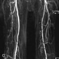
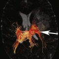
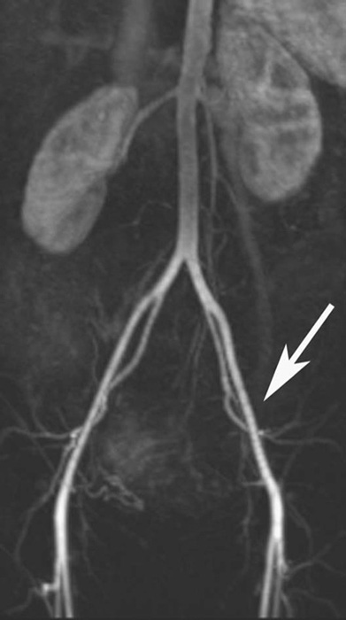
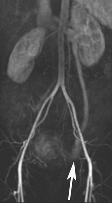
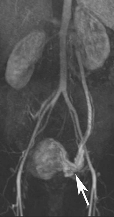
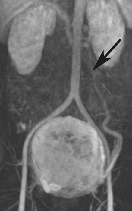
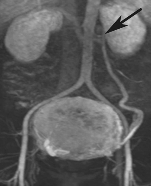
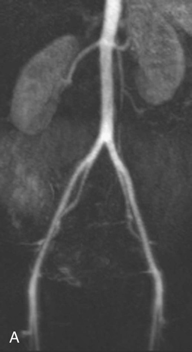
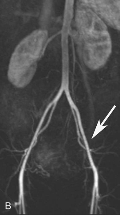
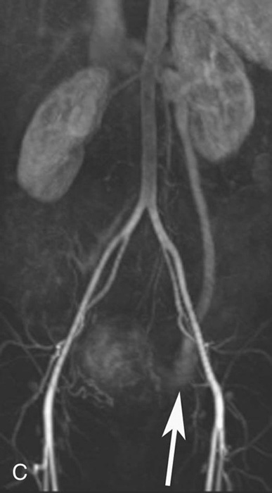
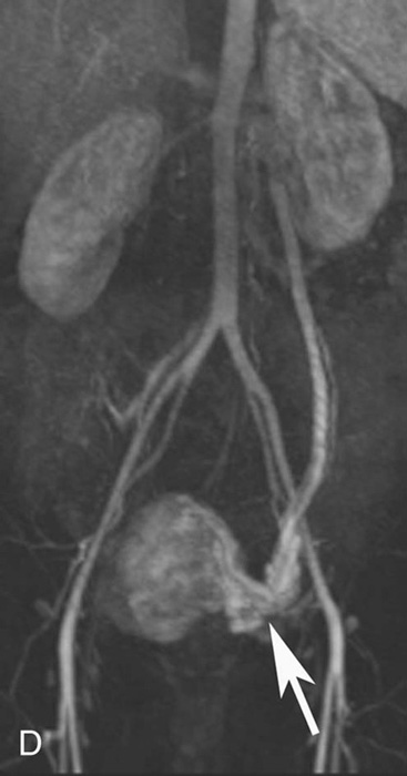
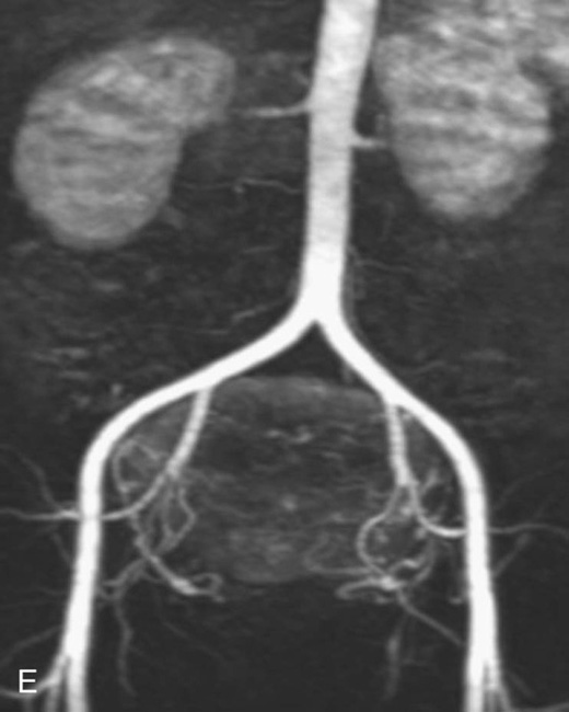
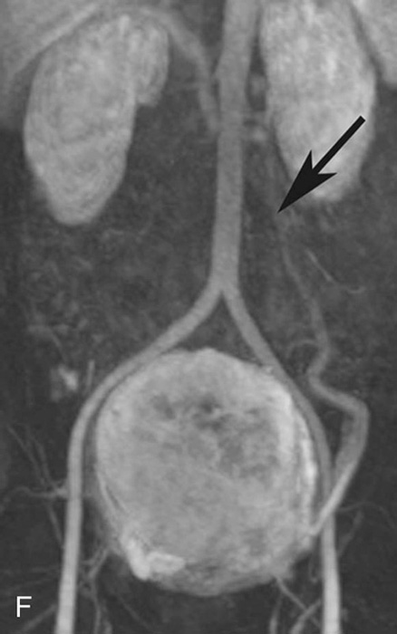
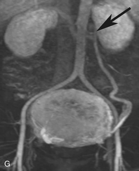
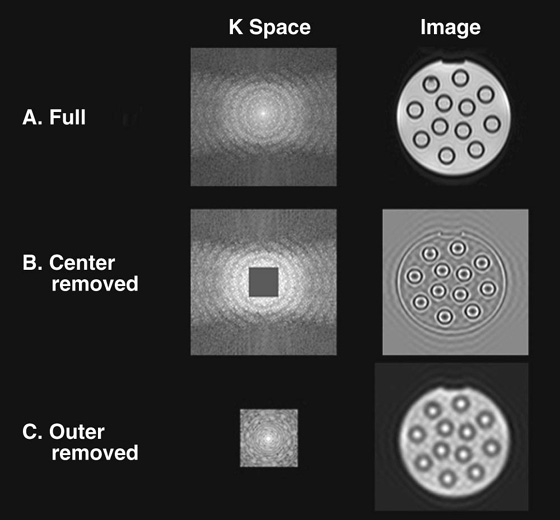
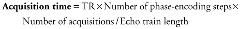
Chapter 12 Kimball L. Christianson, Allen W. Song, Elmar M. Merkle, and Charles Y. Kim 1. What magnetic resonance imaging technique is being used here? 2. What is the diagnosis in the top row of images? What is the diagnosis in the bottom row of images (different patient)? 3. How is this technique useful in interpreting the pathology in these two cases? 4. What does time-resolved imaging mean? FIGURES 1A THROUGH 1D. Time-resolved magnetic resonance angiography (MRA) with interleaved stochastic technique (TWIST) sequence. A through D sequentially show how the contrast flows in a retrograde fashion in the left gonadal vein. The leading edge of the bolus is annotated with white arrows in B, C, and D. FIGURES 1E THROUGH 1G. Time-resolved MRA TWIST sequence. These images also demonstrate a leading edge (also annotated with black arrows in F and G) demonstrating flow in the opposite and correct direction toward the left renal vein. 1. Time-resolved contrast-enhanced MRA. 2. The top row of images demonstrates a dilated left gonadal vein with retrograde flow of contrast in the left ovarian vein, which can be seen with pelvic congestion syndrome. The bottom row of images demonstrates a dilated left gonadal vein with normal direction of blood flow towards the left renal vein. 3. The direction of flow is crucial to making the diagnosis in this case. Static imaging would demonstrate a dilated left gonadal vein in both cases; however, time-resolved imaging with its high temporal resolution allows visualization of the direction the blood is flowing. 4. Time-resolved imaging is the rapid sequential acquisition of images so that the blood flow dynamics (or any other nonstatic property) can be visualized in a real-time manner. When performing time-resolved imaging, there is usually a localized area of interest. A three-dimensional volumetric slab of the area of interest is imaged numerous times during a contrast bolus with very fast acquisitions and very high temporal resolution, with some sacrifice of spatial resolution. Time-resolved magnetic resonance angiography (MRA) can be a helpful tool in making the diagnosis of pelvic congestion syndrome (PCS). PCS is a cause of chronic pelvic pain currently thought to be due to incompetent valves within the ovarian veins that result in reflux of blood and symptoms of congestion and a feeling of fullness in the pelvis. Static imaging, such as computed tomography (CT) and conventional MRA, is limited in evaluation of PCS, as stated in Answer 3 above. The gold standard in diagnosing PCS is angiography. However, time-resolved MRA is a very good alternative because it is noninvasive and does not require ionizing radiation. Endovascular therapy of PCS can be performed by embolization of the ovarian veins using coils or sclerosant.1 Many of the modalities that are used in radiology, including plain films, CT, and much of magnetic resonance imaging (MRI), involve static imaging. Although static images with high resolution are obviously vital to making many diagnoses, sometimes temporal resolution is needed in order to accurately make a diagnosis. Modalities such as ultrasound and fluoroscopy have a high temporal resolution, allowing dynamic imaging to be performed. With the development of time-resolved techniques, MRI now has the capability to perform dynamic imaging. Time-resolved MRA applies very fast imaging techniques that enable multiple acquisitions during a single contrast bolus, thereby offering information on the dynamics of contrast enhancement and actual blood flow dynamics. This is made possible by techniques that have a very rapid image acquistion while still maintaining sufficient spatial resolution. Time-resolved sequences typically employ three-dimensional (3D) sequences that take about 2 to 5 seconds per acquisition.2 Some examples of the use of time-resolved techniques include arteriovenous malformations, peripheral vascular disease, thoracic veins, and differentiating between the true and false lumen in aortic dissection. Time-resolved MRA offers several advantages over static contrast-enhanced MRA, computed tomographic angiography, and conventional angiography. MRA affords a noninvasive examination without use of ionizing radiation. Time-resolved MRA demonstrates the direction of blood flow, which is helpful to detect abnormal retrograde flow or reflux and is especially helpful for evaluating collateral vessels. The limitations of time-resolved MRA include general MRI contraindications—pacemaker, metallic implants, claustrophobia—as well as limited spatial resolution and nephrogenic systemic fibrosis in patients with renal insufficiency. In order to understand the concepts behind time-resolved MRA, it is important to understand the concept of k-space. K-space is where the raw signal resides; the echoes (i.e., magnetic resonance [MR] signals) received by the receiver coils of the magnet are mapped out as spatial frequencies in the k-space. As such, the k-space units are in inverse distance, and the k-space map and MR image (units in distance) are mutually convertible by the Fourier transformation. The coordinates of k-space are referred to as kx and ky in two dimensions and kx, ky, and kz in three dimensions. The center of k-space contributes more to the majority of signal intensity of the MR image and the peripheral k-space contributes to the fine spatial details. This concept is very important in understanding time-resolved imaging. The filling of k-space can be done in many different ways, which are controlled by the so-called trajectories. Linear k-space trajectories fill k-space one line at a time corresponding to each echo. This linear filling of k-space can be done from one edge of k-space to the next, which is a sequential linear trajectory. Alternatively, the central lines of k-space can be filled first with the more peripheral lines filled subsequently. This is known as linear centric k-space filling (Fig. 1). Inherent in understanding how to decrease acquisition times is having a fundamental understanding of the factors that influence acquisition time: All of these components can be manipulated to decrease acquisition times; however, this chapter focuses primarily on methods that decrease the number of phase-encoding steps and undersample the k-space with unique k-space trajectories. One of the strategies used to decrease the number of phase-encoding steps and therefore decrease acquisitions time is called partial Fourier k-space acquisition. This is accomplished by filling only a part of k-space, and then by either determining the remainder of k-space from the information already obtained (since the complex k-space data has conjugate symmetry) or simply performing zero filling of k-space. For example, the central portion of k-space can be quickly obtained if the periphery of k-space is filled in with zeroes, thus dramatically reducing the number of phase-encoding steps. Also, because all of k-space is filled (central actual data and periphery zeroes), the desired matrix size is still acquired and the spatial resolution interpolated.2
Time-Resolved Contrast-Enhanced Magnetic Resonance Angiography
CASE 1


ANSWERS
CASE 1
Discussion:
Physics Discussion
Related posts:
![]()
Stay updated, free articles. Join our Telegram channel

Full access? Get Clinical Tree


Contrast-Enhanced Magnetic Resonance Angiography