Table 20-1.
Muscles of the Abdominal Wall
| MUSCLE | ORIGIN | INSERTION | NERVE SUPPLY |
|---|---|---|---|
| Rectus abdominis | Crest and symphysis of the pubis | Xiphoid process and fifth to seventh costal cartilages | Branches of the lower thoracic |
| External oblique (obliquus externus abdominis) | Fifth to twelfth ribs | Anterior half of the iliac crest, inguinal ligament, and anterior layer of the sheath of the rectus abdominis | Ventral branches of the lower thoracic |
| Internal oblique (obliquus internus abdominis) | Tenth to twelfth ribs and sheath of the rectus abdominis; some fibers from the inguinal ligament terminate in the falx inguinalis | Iliac fascia deep to the lateral part of the inguinal ligament, the anterior half of the iliac crest, and the lumbar fascia | Lower thoracic |
| Transversus abdominis | Seventh to twelfth costal cartilages, lumbar fascia, iliac crest, and inguinal ligament | Xiphoid cartilage and linea alba and through the falx inguinalis, pubic tubercle, and pecten | Lower thoracic |
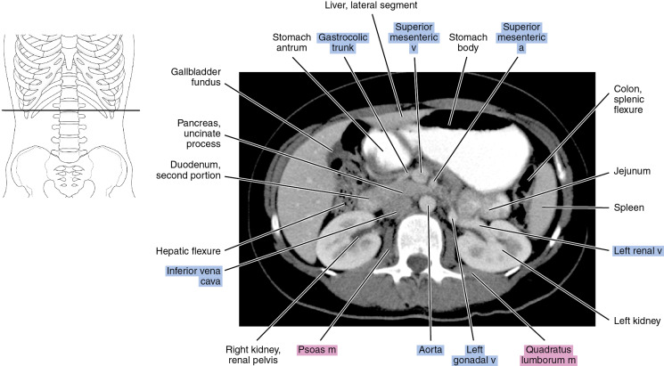
Axial
Figure 20.1.1
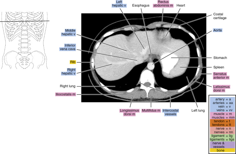
Figure 20.1.2
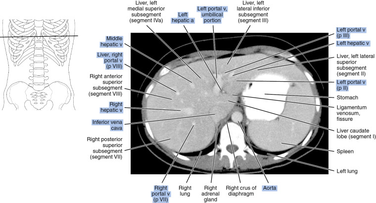
Figure 20.1.3
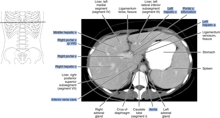
Figure 20.1.4
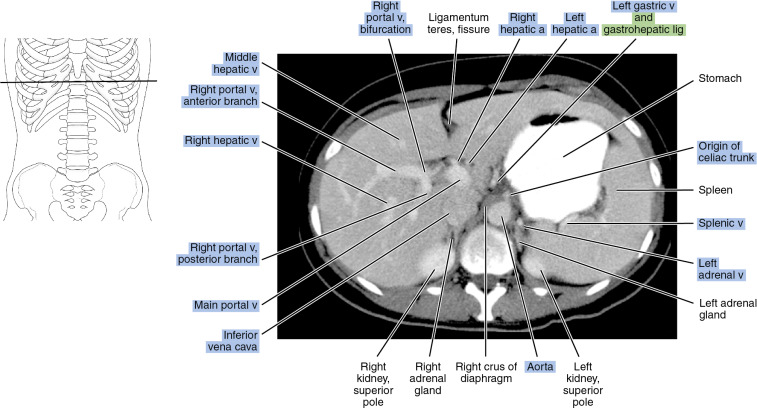
Figure 20.1.5
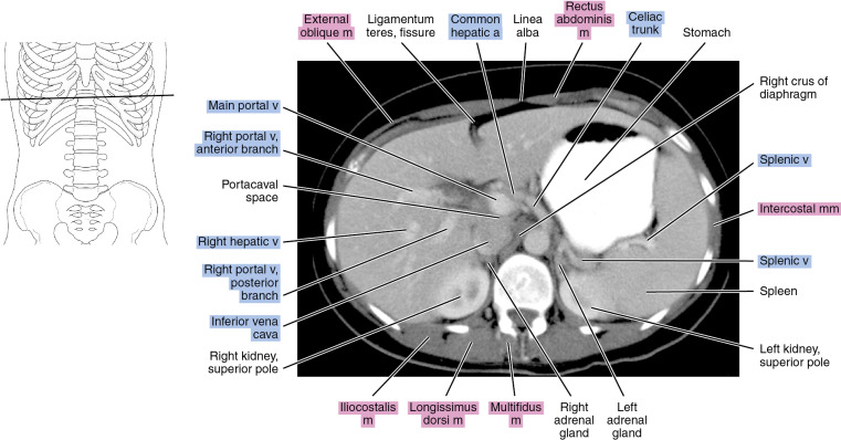
Figure 20.1.6
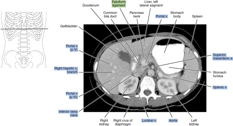
Figure 20.1.7
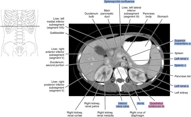
Figure 20.1.8
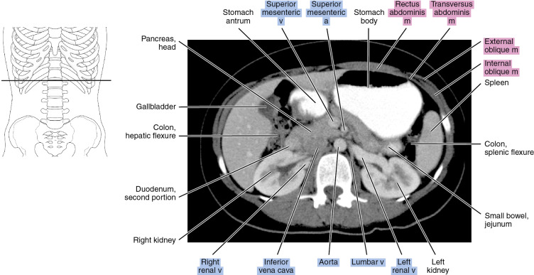
Figure 20.1.9

