Table 8-1.
Muscles of the Hand
| MUSCLE | ORIGIN | INSERTION | NERVE SUPPLY |
|---|---|---|---|
| Palmaris brevis | Ulnar border of the palmar aponeurosis | Deep surface of the skin along the ulnar border of the palm | Superficial branch of ulnar (C8, T1) |
| Abductor pollicis brevis | Palmar surface of the flexor retinaculum, trapezium, and, occasionally, scaphoid | Radial side of the base of the proximal phalanx of the thumb | Recurrent branch of median (C8, T1) |
| Opponens pollicis | Palmar surface of the flexor retinaculum and tubercle of the trapezium | Lateral part of the palmar surface of the shaft of the first metacarpal | Recurrent branch of median (C8, T1) |
| Flexor pollicis brevis | Superficial head: trapezium, adjacent part of the flexor retinaculum, and tendon sheath of the flexor carpi radialis; deep head: trapezoid and capitate | Superficial head: lateral side of the palmar aspect of the base of the proximal phalanx; deep head: into a tendon of the superficial head | Recurrent branch of median and deep branch of ulnar (C8, T1) |
| Adductor pollicis brevis | Carpal head: flexor retinaculum, capitate, bases of the second and third metacarpals; metacarpal head: palmar ridges of the third metacarpal and capsules of the second, third, and fourth metacarpophalangeal articulations | Ulnar side of the palmar aspect of the base of the proximal phalanx of the thumb | Recurrent branch of median (C8, T1) |
| Abductor digiti minimi | Distal half of the pisiform, pisihamate ligament, tendon of the flexor carpi ulnaris, and, frequently, the flexor retinaculum | Two tendons: (1) the ulnar side of the base of the proximal phalanx of the little finger, and (2) the aponeurosis of the extensor tendon of the little finger | Deep palmar division of ulnar (C8, T1) |
| Flexor digiti minimi brevis | Hook of the hamate and adjacent parts of the flexor retinaculum | Ulnar side of the base of the proximal phalanx of the little finger | Superficial or deep palmar branch of ulnar (C8, T1) |
| Opponens digiti minimi | Distal border of the hook of the hamate and adjacent flexor retinaculum | Medial surface of the body and particularly onto the head of the fifth metacarpal | Deep palmar branch of ulnar (C8, T1) |
| Lumbrical | Two lateral lumbricals: radial and palmar sides of the first and second tendons of the flexor digitorum profundus; two medial lumbricals: adjacent side of the second and third tendons, and the third and fourth tendons of the flexor digitorum profundus | Into the radial border of the tendon of the extensor digitorum on the dorsal aspect of the proximal phalanx | Median, lateral two or three lumbricals; ulnar, deep palmar branch, medial one or two lumbricals (C8, T1) |
| Interosseous | Palmar interosseous: anterior border of the shaft of the first, second, fourth, and fifth metacarpals. The first arises near the base and the others arise from three fourths of the shaft of the bone. Dorsal interosseous: adjacent sides of the metacarpal bones in each metacarpal interspace | Into the expansion on the axial side of the corresponding digit. The first palmar interosseous is described frequently as a division of the flexor brevis or adductor pollicis. The first dorsal interosseous usually inserts onto the proximal phalanx. The other three insert into the extensor expansion and proximal phalanx | Deep palmar branch of ulnar (C8, T1) |
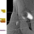
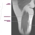
Axial
Figure 8.1.1

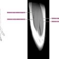
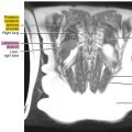
Figure 8.1.2

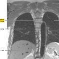
Figure 8.1.3

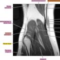
Figure 8.1.4

Figure 8.1.5

Figure 8.1.6