Table 11-1.
Muscles of the Thigh
| MUSCLE | ORIGIN | INSERTION | NERVE SUPPLY |
|---|---|---|---|
| Sartorius | Anterior superior iliac spine and adjacent area below | Medial surface of the tibia; near the tuberosity and neighboring fascia | Femoral |
| Rectus femoris | Straight head: anterior inferior iliac spine; reflected head: posterosuperior surface of the rim of the acetabulum | Through the patellar ligament to the tibial tuberosity | Femoral |
| Vastus lateralis | Shaft of the femur along the anteroinferior margin of the greater trochanter, above the gluteal tuberosity, and the upper half of the linea aspera | Proximal border of the patella, front of the lateral condyle of the tibia and fascia of the leg | Femoral |
| Vastus medialis | Medial lip of the linea aspera and the distal half of the intertrochanteric line, and the aponeurosis of the tendons of insertion of the adductor muscles | Upper two thirds of the medial margin and proximal margin of the patella, medial condyle of the tibia, and investing deep fascia of the leg with the tendons of vastus intermedius, lateralis, and rectus, and through the patellar ligament onto the front of the tibial tuberosity | Femoral |
| Vastus intermedius | Distal half of the lateral margin of the linea aspera and its lateral bifurcation and from the anterolateral part of the shaft of the femur | Proximal margin and deep surface of the patella, aponeurosis of the vastus lateralis, medially and laterally to the tendons of vastus medialis and lateralis, to the patellar ligament and onto the tibial tuberosity | Femoral |
| Gracilis | Medial margin of inferior ramus of the pubis and the pubic end of the inferior ramus of the ischium | By an expanded tendinous process onto the tibia below the medial condyle | Anterior division of the obturator |
| Pectineus | Pectineal line, pectineal fascia, and anterior margin of the obturator sulcus, and from the pubofemoral ligament | Upper half of the pectineal line behind lesser trochanter | Femoral, also from the accessory obturator and/or obturator |
| Adductor longus | Pubic tubercle to symphysis pubis | Middle third of the linea aspera | Anterior division of the obturator; also, occasionally, branch from the femoral |
| Adductor brevis | Medial part of the outer surface of the inferior ramus of the pubis | Distal two thirds of the pectineal line and the upper one third of the linea aspera | Anterior (or posterior) branch of the obturator |
| Adductor magnus | Inferior ramus of the pubis | Medial side of the gluteal ridge and the superior part of the linea aspera by a tendon from the distal three fourths of the linea aspera and the adductor tubercle at the distal end of the medial supracondylar ridge | Posterior branch of the obturator and a branch from the sciatic |
| Biceps femoris | From the lateral lip of the linea aspera of the femur, from the middle of the shaft to the bifurcation of the linea aspera proximal two thirds of the supracondylar ridge, and lateral intermuscular septum | Head of the fibula in front of the apex, partially onto the lateral condyle of the tibia, and into the fascia of the leg | Peroneal part of the sciatic |
Axial
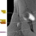
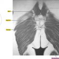
Figure 11.1.1

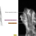
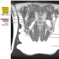
Figure 11.1.2

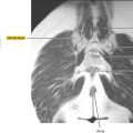
Figure 11.1.3

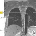
Figure 11.1.4

Figure 11.1.5

Figure 11.1.6

Figure 11.1.7

Figure 11.1.8

Figure 11.1.9

Figure 11.1.10

Figure 11.1.11

Figure 11.1.12

Figure 11.1.13

Figure 11.1.14

Related posts:
Stay updated, free articles. Join our Telegram channel

Full access? Get Clinical Tree


