Table 6-1.
Muscles of the Forearm
| MUSCLE | ORIGIN | INSERTION | NERVE SUPPLY |
|---|---|---|---|
| Anconeus | Posterior surface of the lateral epicondyle, and adjacent capsular ligament of the elbow | Onto the radial side of the olecranon and adjacent shaft of the ulna | Nerve to anconeus (C7, C8, T1) |
| Brachioradialis | Upper two thirds of the lateral epicondylar ridge of the humerus and the anterior surface of the lateral intermuscular septum | Lateral side of the base of the styloid process of the radius | Radial (C5, C6, C7) |
| Extensor carpi radialis longus | Lower third of the lateral epicondylar ridge, lateral intermuscular septum, and extensor tendons from the lateral epicondyle | Lateral aspect of the base of the second metacarpal | Radial (C5, C6, C7) |
| Extensor carpi radialis brevis | Common extensor tendon from the lateral epicondyle, intermuscular septa, and radial collateral ligament of the elbow joint | Dorsal aspect of the base of the third metacarpal | Radial or deep radial (posterior interosseus) (C7, C8) |
| Extensor digitorum | Common extensor tendon | Dorsal digital fibrous expansion covering the dorsum of the proximal phalanx and sides of its base, base of the middle, and distal phalanges | Deep radial (posterior interosseus) (C7, C8) |
| Extensor digiti minimi | Intermuscular septa, overlying fascia, and common extensor tendon | Base of the proximal phalanx of the little finger | Deep radial (posterior interosseus) (C7, C8) |
| Extensor carpi ulnaris | Two heads: (1) distal dorsal aspect of the lateral epicondyle, and (2) proximal three fourths of the dorsal border of the ulna | Onto a tubercle at the base of the fifth metacarpal | Deep radial (posterior interosseus) (C7, C8) |
| Supinator | Dorsal aspect of the lateral epicondyle, ulnar depression distal to the radial notch, and supinator crest | Lateral surface of the radius between the anterior and posterior oblique lines | Deep radial (posterior interosseous) (C5, C6) |
| Abductor pollicis longus | Lateral edge of the proximal part of the middle third of the ulna, adjacent interosseous membrane, dorsal surface of the radius, and, occasionally, the intermuscular septa | Radial side of the ventral aspect of the base of the first metacarpal | Deep radial (posterior interosseus) (C7, C8) |
| Extensor pollicis brevis | Distal end of the middle third of the radius in its dorsal surface, interosseous membrane, and, occasionally, the ulna | Base of the proximal phalanx of the thumb or into the capsule the metacarpophalangeal joint | Deep radial (posterior of interosseus) (C7, C8) |
| Extensor pollicis longus | Middle third of the dorsal surface of the ulna adjacent to the interosseous membrane | Base of the distal phalanx of the thumb | Deep radial (posterior interosseus) (C7, C8) |
| Extensor indicis | Proximal part of the distal third of the posterior surface of the ulna interosseous membrane | Dorsal aponeurosis on the ulnar side of the index finger, adjacent to the base of the proximal phalanx | Deep radial (posterior interosseus) (C7, C8) |
| Pronator teres | Two heads: (1) humeral head (superior half of the ventral surface of the medial epicondyle), and (2) ulnar head (medial border of the coronoid process) | Onto the middle third of lateral surface of the radius | Median (C6, C7) |
| Flexor carpi radialis | Medial epicondyle of the humerus | Base of the second metacarpal and usually, base of the third metacarpal | Median (C6, C7) |
| Palmaris longus | Medial epicondyle | Flexor retinaculum and palmar aponeurosis | Median (C7, C8) |
| Flexor carpi ulnaris | Two heads: (1) medial epicondyle, and (2) medial side of the olecranon, upper two thirds of the dorsal border of the ulna | Primarily onto the pisiform | Ulnar (C7, C8) |
| Flexor digitorum superficialis | Two heads: (1) ulnar (ventral surface of the medial epicondyle, ulnar collateral ligament, ulnar tuberosity, medial border of coronoid process), and (2) radial (anterior oblique line and ventral border below the radial oblique line) | Ventral surface of the shaft of the middle phalanx of each finger | Median (C7, C8, T1) |
| Flexor digitorum profundus | Proximal three fourths of the medial and anterior surface of the ulna and interosseous membrane | Bases of the distal phalanges of the second to fifth digits | Median, anterior interosseous branch (C8, T1) |
| Flexor pollicis longus | Ventral surface of the radius, oblique line, and adjacent interosseus membrane | Base of the distal phalanx of the thumb | Median, anterior interosseous branch (C8, T1) |
| Pronator quadratus | Medial side, ventral surface of the distal fourth of the ulna | Distal quarter of the ventral surface of the radius | Median, anterior interosseous branch (C8, T1) |
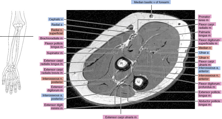
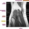
Axial
Figure 6.1.1

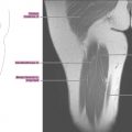
Figure 6.1.2

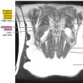
Figure 6.1.3

Figure 6.1.4