and Clarisse Dromain2
(1)
Department of Radiology, San Giovanni Hospital, Roma, Italy
(2)
Department of Radiology, Institut de Cancerologie Gustav Roussy, VilleJuif – Paris, France
Abstract
MR defecography is a study of the pelvis composed by a morphologic study and by a dynamic study dedicated to the functional pelvic floor disease. MR protocol requires patient preparation (rectal cleansing by means of enema) and rectal distension by means of sonographic gel injected through a rectal tube.
Defecography, MR
MR defecography is a study of the pelvis composed by a morphologic study and by a dynamic study dedicated to the functional pelvic floor disease.
MR protocol requires patient preparation (rectal cleansing by means of enema) and rectal distension by means of sonographic gel injected through a rectal tube.
Phase array is used for signal reception. Patient is positioned in supine position.
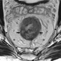
Morphologic study consists in high-resolution T2-weighted sequences acquired on axial, coronal, and sagittal planes in order to study morphologic features of pelvic floor and sphincter complex.
Dynamic study is based on the acquisition of dynamic high-contrast high temporal resolution sequences, fast imaging with steady-state precession (FISP, balanced FFE, FIESTA) sequence. Single-slice sagittal FISP sequence with high temporal resolution is acquired continuously over 40 s time period at rest and during patient straining and defecation.
Descending Perineum Syndrome
Descending perineum syndrome refers to a condition where the perineum descends below the bony outlet of the pelvis during straining and defecation due to weakness of the pelvic floor muscle and excessive straining during defecation. Descending perineum syndrome can be complicated by protrusion of pelvic organs (rectal prolapse and rectocele, colpocele, enterocele, and cystocele) leading to several symptoms including chronic constipation due to “obstructed defecation” (secondary to rectal prolapse and rectocele).< div class='tao-gold-member'>Only gold members can continue reading. Log In or Register to continue

Stay updated, free articles. Join our Telegram channel

Full access? Get Clinical Tree


