and Sofia Gourtsoyianni2
(1)
UOC Radiologia BR, Azienda Ospedaliera Universitaria Integrata Verona, Verona, Italy
(2)
Imaging 2, Level 1, Lambeth Wing St Thomas’ Hospital, London, UK
Diffusion-Weighted Imaging (Focal Liver Lesions)
Diffusion-weighted MR imaging (DW MRI) enables qualitative, by visual assessment, and quantitative assessment of tissue diffusivity, by means of apparent diffusion coefficient (ADC) measurements, without the use of gadolinium chelates, which makes it a highly attractive technique, particularly in patients at risk for nephrogenic systemic fibrosis.
DW MRI is used with great success for the detection and characterisation of focal liver lesions and their differentiation (benign from malignant) in combination with conventional MR sequences. It also provides information regarding tumour treatment response assessment by monitoring change in ADC values of lesions (expressed in 10-3 mm2/s).
Hypercellular tissues, such as tumours or abscesses, demonstrate restricted diffusion (high signal intensity) on higher b value images and lower ADC values. On the other hand, cystic or necrotic lesions show a greater degree of signal attenuation, demonstrating low signal intensity on higher b value diffusion images, and return higher ADC values. One has to keep in mind that lesions may appear to show restricted diffusion due to their long T2-relaxation time as well, when correlating high b value images with corresponding ADC map (lesions show high signal intensity on both). This phenomenon is called T2 shine-through and may be observed in the normal gallbladder, cystic lesions and haemangiomas (pathognomonic). Generally ADC values tend to be lower than those of normal liver parenchyma in malignant tumours and higher in benign liver lesions.
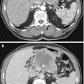
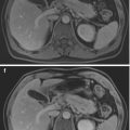
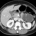
Dorsal Agenesis of the Pancreas
Partial agenesis of the pancreas is an extremely rare malformation, which more often involves the dorsal pancreas. It results from an embryological failure of the dorsal pancreatic bud to form the body and tail of the pancreas. Dorsal agenesis can be isolated or part of heterotaxia syndromes.

Stay updated, free articles. Join our Telegram channel

Full access? Get Clinical Tree


