div style=”display:none;”>
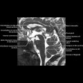
Deep Cerebral Veins
Main Text
TERMINOLOGY
Abbreviations
IMAGING ANATOMY
Overview
Deep Cerebral Veins


div style=”display:none;”>
Deep Cerebral Veins
Main Text
TERMINOLOGY
Abbreviations
IMAGING ANATOMY
Overview