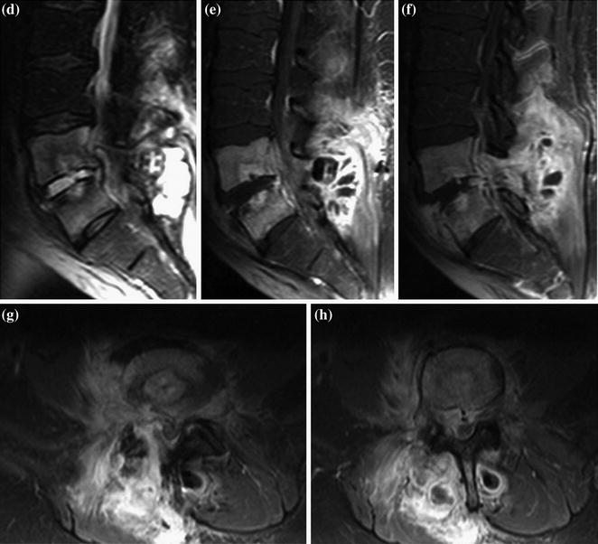
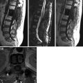
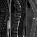
Fig. 1
a–h. SE T1 (a–b), FSE fat sat T2 (c–d) sagittal, CE fat sat SE T1sagittal (e–f) and axial (g–h). Severe spondylodiscitis (T1 hypointensity a–b, T2 hyperintensity c–d) CE of disk, spongiosa, paravertebral soft tissue and right psoas muscle (e–h). Intracanal expansion and soft tissue micro-abscesses along surgical breach
< div class='tao-gold-member'>
Only gold members can continue reading. Log In or Register to continue
Related posts:

Stay updated, free articles. Join our Telegram channel

Full access? Get Clinical Tree


