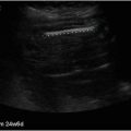
Fig. 4.1
Normal sonographic anatomy of the skin
Glabrous Skin (i.e., Palms and Soles)
Epidermis: bilaminar and thick bright hyperechoic layer due to more prominent keratin content.
Pathology
Epidermal Cyst
Causes: implantation of epidermal elements in the dermis and/or hypodermis.
Ultrasound: The appearance may vary. If the cyst is intact, patients present with a well-defined, round or oval shaped, anechoic structure located in the dermis and/or hypodermis with posterior acoustic enhancement artifact (Fig. 4.2a). An anechoic connecting tract to the subepidermal region called “punctum ” may also be detected. Giant epidermal cysts can show as hypoechoic with anechoic filiform bands due to compacted keratin deposits and an increased presence of cholesterol crystals; this appearance has been called “pseudotestes appearance ” due to their resemblance to the sonographic pattern of the testes. Inflamed cysts may show hyperechoic material. They are larger in size and have increased peripheral vascularity. When the cyst is ruptured, the cyst becomes irregular and the hypoechoic keratinous material can be seen in the periphery, usually generating a “foreign body-like” reaction [4, 7–9] Fig. 4.2b.

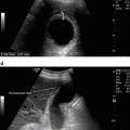
Fig. 4.2
(a) (Top). Epidermal cyst , intact. Hypoechoic oval shaped structure (*) in the dermis and hypodermis with posterior acoustic enhancement artifact (arrows). (b) (Bottom). Ruptured. On color Doppler the cyst (*) presents inflammation and there is spreading of the keratinous material (o) in the right aspect of the image. (d = dermis; h = hypodermis)
Key Sonographic Sign: The posterior acoustic enhancement artifact.
Pilonidal Cyst
Causes : Hairy individuals exposed to chronic friction in the intergluteal region.
Ultrasound : A nest of hyperechoic linear tracts that correspond to fragments of hair tracts trapped in the dermis and hypodermis, surrounded by hypoechoic inflammatory tissue (Fig. 4.3). Commonly, these cysts are connected to the base of widened regional dermal hair follicles. On color Doppler they often show hypervascularity in the periphery due to inflammation and/or infection, in the latter case this is a pilonidal abscess. The aim of the ultrasound study is to show the nature, actual axis and extent of the cyst, which may support the surgical planning [1, 2, 10, 11].

Fig. 4.3
Pilonidal cyst . Notice the hypoechoic structure (*) with fragments of hair tracts (arrowheads) involving dermis and hypodermis. (d = dermis; h = hypodermis)
Key Sonographic Sign: Hyperechoic linear tracts corresponding to the hair tract fragments.
Pilomatrixoma
Synonyms: Calcifying epithelioma of Malherbe or pilomatricoma .
Causes: This is a hair matrix derived tumor most frequently seen in children and young adults and located in the face and extremities. The rate of clinical diagnostic error reported is up to 56 %, therefore ultrasound has an important role in their diagnosis.
Ultrasound: The “target type” of appearance is the most frequent form of presentation and shows as a dermal and hypodermal nodule with a hypoechoic rim and an hyperechoic center with multiple hyperechoic dots corresponding to the calcium deposits. The degree of calcification of these tumors may vary and in cases with large deposits of calcium, a posterior acoustic shadowing may be detected (Fig. 4.4). The presence of blood flow may be variable, going from hypo to hypervascularity with slow flow arterial and venous vessels in the center an d periphery. Rarely, pilomatrixomas can show hypervascularity and clinically mimic a vascular tumor. Another form of presentation is the cystic Pilomatrixoma due to internal bleeding, which appears as an anechoic cystic structure with an eccentric hypoechoic solid nodule with hyperechoic spots [10, 12–15].

Fig. 4.4
Pilomatrixoma . Target type of nodule with hypoechoic rim (r) and hyperechoic center (c) that shows hyperechoic spots that correspond to calcium deposits (arrows). Notice the posterior acoustic shadowing due to the calcium deposits (arrowheads). (d = dermis; h = hypodermis)
Key Sonographic Sign: Hyperechoic spots due to calcifications.
Dermatofibroma
Synonyms: Fibrous histiocytoma and histiocytoma cutis.
Causes: This is a fib romatous tumor most commonly seen in the trunk and extremities of females. It has been postulated to be a reactive reaction to trauma such as an insect bite and also a benign neoplastic disorder.
Ultrasound: An ill-defined hypoechoic and heterogeneous solid dermal structure usually with distortion and enlargement of the regional hair follicles (Fig. 4.5). Vascularity may be variable, commonly with thin, slow flow arterial and venous vessels [10].

Fig. 4.5
Dermatofibroma . Ill-defined hypoechoic lesion (*) that distorts the regional hair follicles. (d = dermis; h = hypodermis)
Key Sonographic Sign: Distortion o f the lesional hair follicles.
Lipoma
Causes: A benign proliferation of mature adipose cells. According to the tissue that accompanies these fatty cells the lipoma may be called fibrolipoma (fibrous tissue) or angiolipoma (capillary vessels). Lipomas are the most common soft tissue tumor and they are usually single, however they can be multiple.
Ultrasound: They show as round or oval shaped hypodermal structures that tend to follow the axis of the skin layers and present hyperechoic septa. Their echogenicity also changes according to the type of tissue that is attached to the adipose cells. Thus, fibrolipomas tend to show hypoechogenicity and angiolipomas present hyperechogenicity. The sonographer should be aware of risky locations of lipomas such as the neck or temple regions where thick vessels or nerves can be located in the vicinity. Occasionally, lipomas can be located underneath the epicranius muscle in the frontal region, an entity called subgaleal lipoma, which may clinically simulate an epidermal cyst or an exostosis. Frequently, lipomas are hypovascular, and the presence of heterogenicity and hypervascularity within the tumor support the suspicion of a malignant transformation [10, 16–18].
Key Sonographic Sign: Structure that follows the axis of the skin layers. Caution; lipoma s are usually located in the more superficial areas of the skin. If a lesion is scanned deeper next to the muscle, the patient should be referred for further workups as malignancies can present this way.
Malignant Tumors
Non-melanoma Skin Cancer (NMSC)
General Concepts : NMSC is the most frequent cancer in hum an beings and frequently affects highly sun-exposed areas of the body such as the face. NMSC is conformed by basal cell (BCC) and squamous cell carcinoma (SCC), BCC being the most common type. Although NMSC is rarely lethal, it is highly disfiguring nevertheless. The ultrasound–histology correlation of the depth of BCC has been reported as excellent.
Ultrasound: They present as hypoechoic and/or heterogeneous oval shaped lesions with irregular or lobulated borders, commonly located in the dermis. BCC typically shows hyperechoic spots within the tumor and the extent of the presence of these spots seems to be correlated with the high or low risk of recurrence of histologic subtypes (Fig. 4.6). BCCs with a higher number of hyperechoic spots (≥7) have been reported to correlate well with more aggressive histologic subtypes such as micronodular, morpheiform, sclerosing, infiltrating, and metatypical subtypes [19–24]. SCCs do not present hyperechoic spots and tend to be more infiltrating than BCCs, more easily extending into the underlying muscle or cartilage. On color Doppler, NMSC presents slow flow vascularity within the lesions with arterial and venous vessels; SCC is usually more hypervascular than BCC [22].

Fig. 4.6
Basal cell carcinoma. Hypoechoic oval shaped structure (*) with hyperechoic spots (between markers) that involves dermis and hypodermis. (d = dermis; h = hypodermis)
Objective of the Examination: To support the detection of the NMSC lesion , define its extent, including depth, support the p rediction of type (BCC or SCC) and also the subtype (high or low risk of recurrence BCC) as well as demonstrate the involvement of deeper layers.
Key Sonographic Sign: Hyperechoic spots for BCC.
Melanoma
General Concepts: Melanoma is the most lethal form of skin cancer and is usually a hyperpigmented lesion. However, there are amelanotic melanomas where the pigment is hidden in the tumor. Melanomas tend to rapidly spread into nodal or extra-nodal metastases.
Primary Tumor
General Considerations: In spite of the fact that melanoma is not the most frequent type of skin cancer, it is the most aggressive type. The recurrence of melanoma has been reported to be up to 46.1 % in lesions of the head and neck region. Importantly, the prognosis of the patient is provided by the depth of the tumor invasion through the Breslow index. This is a histologic measurement of how deep the tumor is. However, ultrasound can also measure the extent of the tumor by providing a sonographic Breslow index. Ultrasound can help to differentiate melanomas that measure < or >1 mm; this can support relevant clinical decisions such as the noninvasive assessment of the free margins for surgery, the size of the excision and the performance of a sentinel node procedure (indicated in melanomas measuring >1 mm).
Ultrasound: They tend to show as well-defined, hypoechoic and or heterogeneous oval shaped and usually fusiform structures located in the dermis and/or hypodermis. Frequently, they show hypervascularity due to their angiogenic power and also may extend to deeper layers (Fig. 4.7a, b). Using color Doppler and contrast enhanced ultrasound studies, it has been reported that the degree of maligna ncy usually correlates well with the degree of vascularity [25–32].

Fig. 4.7
(a) (Top). Melanoma . Grey scale shows hypoechoic oval shaped mass (*) involving dermis and hypodermis (h). (b) (Bottom). On color Doppler prominent vascularity is detected within the mass (*)
Locoregional Staging
General Concepts: Ultrasound is more sensitive than clinical palpation for the diagnosis of metastasis. Ultrasound has been proven useful for diagnosing extranodal and nodal metastasis [25].
Extranodal Metastases
These can be separated into satellites (<2 cm from the primary tumor) and in-transit (≥2 cm from the primary tumor). Satellite or in-transit metastases often appear as hypoechoic nodules with irregular borders and increased echogenicity of the surrounding hypodermis due to perilesional edema. Metastases can show anechoic areas, most probably due to increased transmission of the sou nd in hypercellular regions. Hypervascularity through irregular, thick and tortuous vessels may also be seen [33–40].
Nodal Metastases
Lymph node criteria for benignancy and malignancy are shown in Table 4.1. This staging follows the lymphatic drainage according to the anatomical location of the tumor. Additionally, ultrasound can support a percutaneous fine-needle aspiration (FNA) cytology (FNAC) or biopsy (FNAB) of the lymph nodes [41–44].
Table 4.1
Color Doppler ultrasound o f benign and malignant lymph nodes
Ultrasound morphology | Benign | Malignant |
|---|---|---|
Shape | Oval | Round |
Center | Hyperechoic | Hypoechoic/anechoic |
Cortex thickening | Diffuse | Nodular, eccentric |
Ratio longitudinal/transverse | >2 | <2 |
Vascularity | Regular, central | Irregular, peripheral |
Cortex vessels | Absent/few | Present/prominent |
Key Sonographic Sign: Fusiform hypoechoic and hypervascular structure in correlation with a hyperpigmented lesion.
Vascular Anomalies
General Concepts: These usually affect the pediatric population and are common lesions for referral. The most common vascular anomalies that are sent for ultrasound examination are hemangiomas and vascular malformations. These have different origins, evolution, treatment and prognosis. Thus, sonographic support in the differentiation of these two entities can be critical f or proper management.
Hemangiomas
General Concepts: These are the most common soft tissue lesions in infancy and present rapid growth during the first year and then they start a slow regression period.
Ultrasound: The sonographic appearance of hemangioma varies according to the phase. In the proliferative phase, they show as an ill-defined hypoechoic mass with hypervascularity that includes arterial and venous vessels and some arteriovenous shunts. In the partial regression phase, the echogenicity becomes mixed and the lesion presents hypoechoic and hyperechoic regions (Fig. 4.8a, b). The blood flow also decreases in this phase. In the total regression phase, the hemangioma turns to hyperechoic and hypovascular. Ultrasound may support the follow-up of the treatment and provide anatomic information on the current extent, phase, and involvement of deeper layers or structures such as muscle, cartilage, glands, or the ocular globe. Also ultrasound can be used for monitoring treatment such as propranolol [45–48].

Fig. 4.8
(a) (Top). Hemangioma partial regression phase. Ill-defined heterogeneous structure (*) that involves dermis and hypodermis. (b) (Bottom). 3D power angio reconstruction demonstrates areas with prominent vascularity (*) within the lesion and areas without detectable flow
Key Sonographic Sign: Ill-defined hypoechoic or heterogeneous hypervascular structure that changes in its echogenicity and degree of vascularity over time.
Vascular Malformations
General Concepts: These are errors of morphogenesis and do not comprise an actual tumor. They can be separated according to the type of vessel (arterial, venous, capillary, or lymphatic) and to the high (arterial or arteriovenous) or low flow (venous, capillary, or lymphatic).
Ultrasound: A sonographic sign is the presence of a nest of anechoic tubular structures or lacunar spaces. Thus, on color Doppler ultrasound with spectral curve analysis, they can demonstrate their arterial (with systolic and diastolic component), venous (monophasic), or arteriovenous (to and fro flow) features (Fig. 4.9a, b). Capillary vascular malformations can show epidermal thickening, decreased echogenicity of the upper dermis or increased echogenicity of the hypodermis and lack of blood flow due to their being very slow velocity vessels. Ultrasound can also guide a percutaneous sclerosing procedure [45–47].

Fig. 4.9
(a) (Top). High flow arterial vascular malformation . Notice the hypervascular spots in the dermis of the nasal tip. (b) (Bottom). Spectral curve analysis demonstrates arterial flow. (d = dermis; c = nasal cartilage)
Key Sonographic Sign: anechoic tubular and tortuous nest of vessels.
Inflammatory Diseases
Psoriasis
General Concepts: A chronic autoimmune inflammatory disorder that involves the skin, nail, tendons, joints, and bone.
Ultrasound
Skin: psoriatic cutaneous plaques show as thickening of the epidermis and decreased echogenicity of the upper dermis. Occasionally, undulation of the epidermis may also be seen. On color Doppler examination, dermal hypervascularity with slow flow arterial and venous vessels may be detected in the subepidermal region (Fig. 4.10a).

Fig. 4.10


(a) (Top) Psoriasis . Cutaneous plaque shows epidermal and dermal thickening and increased regional blood flow on power Doppler. The vertical white lines are pointing out the normal (left aspect) and abnormal regions (right aspect). (b) (Bottom). Nail involvement demonstrates thickening of the ventral plate (arrow) and the nail bed
Related posts:
Stay updated, free articles. Join our Telegram channel
Full access? Get Clinical Tree
