Diabetic Foot with Gas-Forming Infection
Andrew F. Barnes
Daniel B. Nissman
CLINICAL HISTORY
53-year-old male with history of type 2 diabetes mellitus presents with a non-healing ulcer on the plantar surface of his right foot at the base of the great toe. He also complains of fevers and diffuse erythema of the right foot.
 FIGURE 100A |
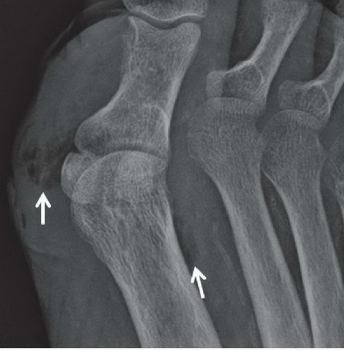 FIGURE 100B |
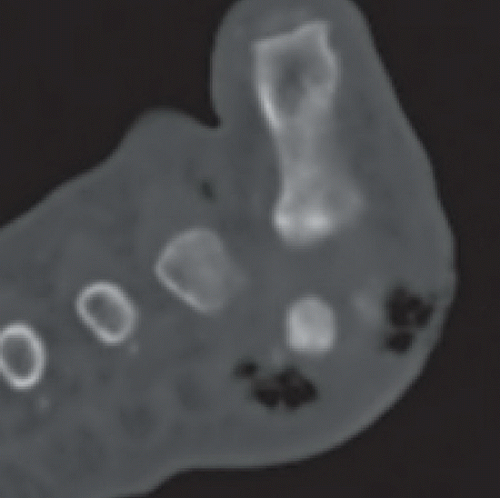 FIGURE 100C |
FINDINGS
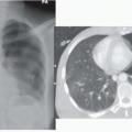
AP and oblique radiographs of the right foot (Figs. 100A and 100B) demonstrate soft tissue edema and subcutaneous gas in the soft tissues about the first metatarsophalangeal (MTP) joint, which also tracks along the first and second metatarsals. The oblique view demonstrates a soft tissue defect subjacent to the first MTP joint. Axial CT of the right foot (Fig. 100C) at the level of the metatarsal heads reveals soft tissue gas about the first MTP joint with an associated superficial cutaneous defect.
Related posts:


Stay updated, free articles. Join our Telegram channel

Full access? Get Clinical Tree


