Diffuse Axonal Injury
James C. Darsie
CLINICAL HISTORY
27-year-old man involved in a high-speed motorcycle-versus-car collision with a poor neurologic examination.
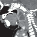
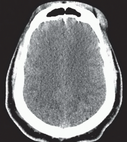 FIGURE 69A |
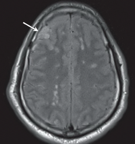 FIGURE 69B |
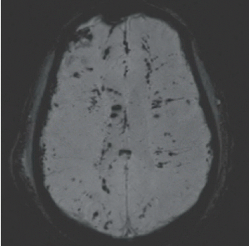 FIGURE 69C |
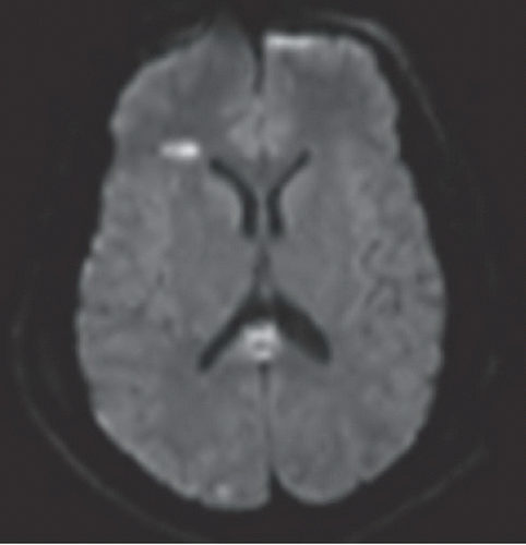 FIGURE 69D |
FINDINGS
Figure 69A: Noncontrast head CT image demonstrates effacement of the cortical sulci, suggesting edema, but preservation of gray-white differentiation. There was also evidence of subarachnoid and intraventricular hemorrhage (not shown), but no definite intraparenchymal hemorrhage is seen. Figures 69B,69C and 69D are images from a subsequent MRI performed 3 days later because of continued poor neurologic examination and clinical suspicion for
traumatic brain injury (TBI). Figure 69B: FLAIR image at a roughly comparable level to Figure 69A shows multiple punctate hyperintensities in the white matter of the centrum semiovale. There is also a more confluent area of cortical hyperintensity in the right frontal lobe, compatible with a cortical contusion. Figure 69C: Susceptibility-weighted imaging (SWI) demonstrates multiple punctate and linear foci of dark signal, most prominently at the gray-white matter junction throughout the cerebrum and corpus callosum. Figure 69D: Diffusion-weighted image (DWI) demonstrates areas of increased signal within the brain and splenium of the corpus callosum that were restricted on the ADC map (not shown). Postcontrast imaging is not shown, but these lesions did not demonstrate contrast enhancement.
traumatic brain injury (TBI). Figure 69B: FLAIR image at a roughly comparable level to Figure 69A shows multiple punctate hyperintensities in the white matter of the centrum semiovale. There is also a more confluent area of cortical hyperintensity in the right frontal lobe, compatible with a cortical contusion. Figure 69C: Susceptibility-weighted imaging (SWI) demonstrates multiple punctate and linear foci of dark signal, most prominently at the gray-white matter junction throughout the cerebrum and corpus callosum. Figure 69D: Diffusion-weighted image (DWI) demonstrates areas of increased signal within the brain and splenium of the corpus callosum that were restricted on the ADC map (not shown). Postcontrast imaging is not shown, but these lesions did not demonstrate contrast enhancement.
Related posts:


Stay updated, free articles. Join our Telegram channel

Full access? Get Clinical Tree


