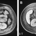
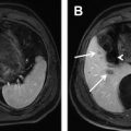
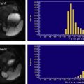
Liver metastases are the most frequently encountered malignant liver lesions in the Western countries. Accurate diagnosis of liver metastases is essential for appropriate management of these patients. Multiple imaging modalities, including ultrasound, CT, positron emission tomography, and MRI, are available for the evaluation of patients with suspected or known liver metastases. Contrast-enhanced MRI has a high accuracy for detection and characterization of liver lesions. Additionally, diffusion-weighted MRI (DWI) has been gaining increasing attention. It is a noncontrast technique that is easy to perform, could be incorporated in routine clinical protocols, and has the potential to provide tissue characterization. This article discusses the basic principles of DWI and discusses its emerging role in the detection of liver metastases in patients with extrahepatic malignancies.
Background of liver metastases
Liver metastases are the most frequently encountered malignant liver lesions in the Western countries, including the United States. In a large German autopsy series of more than 12,000 cases, liver metastases were found to represent 5.0% of cases, followed by hepatocellular carcinoma (3.1%). Many primary neoplasms, including colorectal, breast, kidney, pancreas, and melanoma, can metastasize to the liver. Accurate diagnosis of liver metastases is essential for appropriate management of these patients; patients without evidence of metastases to the liver and extrahepatic sites may benefit from definitive surgical treatment, whereas patients with liver metastases may need to be treated with systemic chemotherapy or segmental hepatectomy, or locoregional therapies such as chemoembolization and radiofrequency ablation. Although patients with liver metastases, especially from colorectal cancer, who meet the criteria for surgical resection have significant improvement in survival, fewer than 25% of patients with disease limited to the liver are candidates for surgical resection. Hence, in patients with primary extrahepatic malignancies, accurate detection of liver metastases is essential.
Role of imaging for detection of liver metastases
Multiple imaging modalities, including ultrasound, CT, positron emission tomography (PET), PET-CT, and MRI are available for evaluating patients with suspected or known liver metastases. Transabdominal ultrasound has a limited sensitivity of 50% to 75% for detecting hepatic lesions, and even lower sensitivity for smaller lesions. The use of contrast-enhanced ultrasound can improve lesion detection and characterization, but the ultrasound contrast is not yet approved by the US Food and Drug Administration (FDA). Furthermore, ultrasound is user-dependent and may require another confirmatory study to characterize liver lesions in many instances.
Multidetector CT (MDCT) is routinely used for evaluating metastatic disease in patients with primary malignancies. It is widely available, relatively inexpensive, and user independent. Multiphasic contrast-enhanced CT with acquisition in both the arterial (20 to 30 seconds after contrast injection) and portal venous phases (70 to 90 seconds after contrast administration) of enhancement has been shown to optimize detection of hypervascular and hypovascular liver metastases, respectively. Limitations of multiphasic CT include exposure to ionizing radiation, and the risk of contrast induced nephropathy from exposure to iodinated contrast media. Another limitation of CT is its inability to definitively characterize subcentimeter lesions. These lesions are reported as “too small to accurately characterize,” even in patients with known primary malignancies.
Fluorodeoxyglucose (FDG)-PET and PET-CT have been used increasingly in the evaluation of metastatic disease in patients with primary malignancies. Studies have shown high sensitivity and specificity for detection of liver metastases. Truant and colleagues reported a similar sensitivity of 76% for both CT and FDG-PET. Ogunbiyi and colleagues reported that FDG-PET had a high sensitivity (95%) and specificity (100%) for detecting liver metastases, and that it was superior to that of the CT, which had sensitivity and specificity of 74% and 85%, respectively.
In a meta-analysis that reviewed 61 articles published between 1990 and 2003 to compare CT, PET-CT, and MRI in the diagnosis of colorectal liver metastases, the authors concluded that PET-CT had a significantly higher sensitivity on a per-patient basis but not on a per-lesion basis. PET has also been shown to be superior to CT for detecting extrahepatic metastases. Lai and colleagues found previously unsuspected extrahepatic disease, predominantly involving the celiac lymph nodes, in 32% patients scheduled for hepatic metastasectomy. Sahani and colleagues reported that FDG-PET identified extrahepatic disease in 9 of the 34 patients involved in their study. However, limitations of PET-CT include radiation exposure, cost, and limited availability.
CT and ultrasound allow evaluation of liver lesions based on a single physical parameter, such as attenuation and echotexture, respectively. In contrast, MRI provides an unique ability to interrogate multiple parameters like T1, T2, diffusion, and enhancement patterns of focal liver lesions, enabling accurate liver lesion detection and characterization ( Table 1 ). Three-dimensional T1-weighted fat-suppressed gradient-recalled echo sequence performed before and after intravenous contrast administration is the workhorse of the liver MR examination. Multiphasic acquisition after injection of extracellular gadolinium-based contrast agents during the hepatic arterial, portal venous, and equilibrium phases can be obtained without radiation exposure and has been shown to have high accuracy in the detection and characterization of liver lesions. MRI with the use of intravenous gadolinium contrast agents was found to have higher accuracy (98% vs 86% and 66%, respectively) than contrast-enhanced PET-CT and unenhanced PET-CT for detecting colorectal metastases. Liver-specific contrast agents, including hepatobiliary agents such as gadolinium-ethoxybenzyl (Gd-EOB-DTPA, Eovist or Primovist, Bayer Healthcare Pharmaceuticals, Montville, NJ, USA) and gadobenate dimeglumine (Gd-BOPTA, MultiHance, Bracco Diagnostics Inc, Princeton, NJ, USA), and reticuloendothelial agents such as superparamagnetic iron oxide particles (SPIO, Feridex, Bayer Healthcare Pharmaceuticals, Montville, NJ, USA), have shown considerable promise in further improving the sensitivity and specificity of MRI for diagnosis and characterization of focal liver lesions. However, SPIO contrast was recently withdrawn from the market in the United States.
| Sequence | Acquisition Plane | TR/TE | Slice Thickness (mm)/gap (%) | Matrix |
|---|---|---|---|---|
| T1 GRE in and out of phase | Axial | 188/2.2–4.4 | 8/20 | 178 × 256 |
| T2 TSE fat-suppressed | Axial | ∼2000–4000/85 | 8/20 | 192 × 256 |
| Single shot T2 TSE non fat suppressed | Coronal | 90 – infinity/65 | 4/20 | 256 × 256 |
| Fat-suppressed SS EPI diffusion a (breath-hold or respiratory triggered) | Axial | 1600–2000 (BH)-1 respiratory cycle (RT)/minimum | 6 (RT)–7 (BH)/20 | 144 × 192 |
| Three-dimensional T1 fat-suppressed GRE Pre– and 3 post–contrast acquisitions (dual arterial, portal venous [60 s], and equilibrium [180 s] phases) | Axial | 3.5/1.6 | 2.1–2.5/NA | 256 × 256 (interpolated) |
a b Values: 0, 50, 500 s/mm 2 (BH) or 0, 50, 500, 1000 s/mm 2 (RT).
Drawbacks of MRI include limited availability, higher cost, and longer examination time that requires patients to hold their breath during many acquisitions. Nephrogenic systemic fibrosis is a rare and potentially life-threatening condition that has been recently reported in patients with advanced renal failure receiving gadolinium-based contrast agents. Consequently, extreme caution is advised in administering gadolinium-based contrast material in patients with severely impaired renal function (glomerular filtration rate <30 mL/min/1.73 m 2 ). Therefore, noncontrast MR techniques, such as diffusion-weighted imaging (DWI), are attractive in this patient population.
DWI acquisition, processing, and interpretation
Principles of DWI
As a result of recent advances in MR technology, DWI is now feasible outside the brain with improved image quality and promising results. This noncontrast technique is easy to implement in clinical practice as it can be added to the existing protocols without significant time penalty.
Diffusion-weighted imaging quantifies thermally induced motion of water molecules known as Brownian motion in tissues. The apparent diffusion coefficient (ADC) calculation has been used to quantify combined effects of capillary perfusion and tissue diffusion.
The ADC is calculated by performing a monoexponential fit to the relationship between the measured signal intensity (in logarithmic scale) and the b values as follows:
Stay updated, free articles. Join our Telegram channel

Full access? Get Clinical Tree


