Disorders of the Cardiac Rhythm


CLINICAL OVERVIEW


Cardiac imaging has a rapidly increasing role in the evaluation and management of patients with cardiac rhythm disorders. Atrial dysrhythmias are among the most common indications for cardiac imaging studies. Atrial fibrillation (AF) is a well-established risk factor for ischemic stroke, arterial thromboembolism, heart failure (HF), and dementia. AF is present in approximately 3% of the adult population. Imaging the left atrium (LA) plays a critical role in AF management. Ventricular dysrhythmias, although less common, are associated with significant morbidity and mortality in patients with heart disease. Scar tissue after myocardial infarction (MI) and in other cardiomyopathies is an arrhythmogenic substrate for the formation of ventricular tachycardia (VT).


DIAGNOSTIC STRATEGY AND THERAPEUTIC IMPLICATIONS


Atrial Dysrhythmias


Left Atrial Morphology and Function


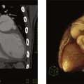
Among its other roles, the LA acts as a conduit and reservoir for blood returning to the heart from pulmonary circulation. Blood enters the LA posteriorly through the four pulmonary veins (PVs) and then flows anteriorly toward the mitral valve. The LA is separated from the right atrium by the interatrial septum (IAS), which is connected anteriorly to the aortic root ( Fig. 11.1 ). LA volumes can be measured by transthoracic echocardiography (TTE), cardiac computerized tomography (CCT) and cardiovascular magnetic resonance (CMR). Studies have shown that larger LA volumes are associated with AF recurrence after ablation. The left atrial appendage (LAA) arises from the LA and is an embryological remnant of the primordial LA. Its lateral border is the left lateral ridge, with infoldings of the atrial wall filled with external adipose tissue, separating the LAA from the left superior PV. The LAA is a long, lobed, tubular structure. The identification of the multiple lobes and their direction is paramount when imaging the LAA, both for preprocedural planning and intraprocedural guidance of LAA closure devices, and for ruling out or in the presence of an LAA thrombus. Four typical morphologies are used to describe the LAA: chicken wing (most common), windsock, cauliflower, and cactus ( Fig. 11.2 ). These can be identified in vivo by multiple imaging modalities (including CCT, CMR), transesophageal echocardiography (TEE), and angiography ( Fig. 11.3 ). These different shapes also correlate with varied stroke risks, as the cactus-shaped LAA most correlates with a history of stroke and the chicken wing shape with an absence of stroke history. The LAA is lined with pectinate muscles that form indentations along its inner surface. Occasionally, bulkier pectinate muscles can be confused with thrombi ( Fig. 11.4 ).


Fig. 11.1


Anatomy of the left atrial appendage (LAA). Anatomic relationships of LAA with surrounding structures from (A) an external and (B) an internal perspective. Ao , Aorta; CX , circumflex artery; LDA , left anterior descending artery; LLR , left lateral ridge; LUPV , left upper pulmonary vein; MV , mitral valve.

Adapted from Faletra FF, Saric M, Saw J, et al. Imaging for Patient’s selection and guidance of LAA and ASD percutaneous and surgical closure. JACC Cardiovasc Imaging 2021;14(1):3–21.


Fig. 11.2


Anatomic variants of left atrial appendage (LAA) morphology. Images taken from explanted hearts demonstrating different LAA morphologies. (A) Chicken wing. (B) Windsock. (C) Cauliflower. (D) Cactus.

Adapted from Beigel R, Wunderlich NC, Ho SY, et al. The left atrial appendage: anatomy, function, and noninvasive evaluation. JACC Cardiovasc Imaging . 2014;7(12):1251–1265.


Fig. 11.3


The four different left atrial appendage morphologies as shown by transesophageal echo (top), cine angiography (middle), and three-dimensional computed tomography (bottom). Cauliflower (A–C), windsock (D–F), cactus (G–I), and chicken wing (J–L).

Adapted from Beigel R, Wunderlich NC, Ho SY, et al. The left atrial appendage: anatomy, function, and noninvasive evaluation. JACC Cardiovasc Imaging . 2014;7(12):1251–1265.


Fig. 11.4


Abnormal transesophageal echocardiography (TEE) findings within the left atrial appendage (LAA). (A) The presence of a large pectinate muscle can sometimes be confused for an LAA thrombus. (B) In this case, the pectinate muscle is better defined by three-dimensional (3D) TEE. (C) spontaneous echocardiographic contrast is seen in the LAA. (D) A more echo-dense, amorphous finding consistent with sludge is seen within the LAA ( arrowheads ). Zero-degree (E) and 95-degree (F) views and 3D imaging (G) show a thrombus within the LAA ( arrows ).

Adapted from Beigel R, Wunderlich NC, Ho SY, et al. The left atrial appendage: anatomy, function, and noninvasive evaluation. JACC Cardiovasc Imaging . 2014;7(12):1251–1265.


Left Atrial Thrombi


Patients with a history of AF are more likely to develop thrombus in the LA and LAA due to reduced contractility and increased stasis. This is often associated with reduced LAA emptying velocities on TEE Doppler studies. The LAA normally contracts in patients in sinus rhythm, however, patients with significantly elevated LV end-diastolic pressure may also be at risk for LAA thrombus formation, despite remaining in sinus rhythm. TEE is highly accurate in the detection of LA thrombi with a sensitivity of 93% and specificity of 100% and is considered the imaging reference standard. Using multiplane 2-dimensional TEE, as well as tissue and spectral Doppler, the LA and LAA can be thoroughly examined ( Fig. 11.5 ). Multiple findings in the LAA can represent different stages of thrombus formation ( Figs. 11.4, 11.6, and 11.7 ). Ultrasound contrast agents may be administered during the procedure to differentiate spontaneous echo contrast (smoke) from LAA thrombus ( Fig. 11.8 ). The presence of spontaneous echo contrast correlates with LA thrombus and is associated with reduced blood velocities and increased LA size. An algorithm for using TEE before elective cardioversion is shown in Fig. 11.9 .


Fig. 11.5


Evaluation of left atrial appendage (LAA) thrombus. (A) Common transesophageal echo planes used for examination, (B) three-dimensional reconstruction showing multiple LAA lobes. LV , Left ventricle.

Adapted from Donal E, Galli E, Lederlin M, et al. Multimodality imaging for best dealing with patients in atrial arrhythmias. JACC Cardiovasc Imaging 2019:2245–2261.


Fig. 11.6


Left atrial appendage (LAA) spontaneous echo contrast. Images obtained from a patient before DC cardioversion showing LAA spontaneous echo contrast (smoke).


Fig. 11.7


Left atrial appendage (LAA) thrombus. Trans-esophageal echocardiography (TEE) images obtained from a patient before DC cardioversion showing a large thrombus in the left atrium and LAA.


Fig. 11.8


Ultrasound contrast agents for evaluation of the left atrial appendage (LAA). Transesophageal echo images obtained from a patient before DC cardioversion excluding LAA thrombus.


Fig. 11.9


Algorithm for approach to transesophageal echocardiography (TEE) before cardioversion. LAA , Left atrial appendage.

Adapted from Beigel R, Wunderlich NC, Ho SY, et al. The left atrial appendage: anatomy, function, and noninvasive evaluation. JACC Cardiovasc Imaging . 2014 Dec;7(12):1251–1265.


CCT and CMR are imaging modalities that can be used for the assessment of thrombus in the LA and LAA. When compared to TEE, CCT has a similar ability to detect LAA thrombus, with the caveat that only TEE has been validated with surgical or postmortem anatomical findings. CCT with delayed imaging after contrast injection ensures maximal contrast filling of the LAA and lowers false positive rates ( Figs. 11.10, 11.11 ).


Fig. 11.10


Axial images of the left atrial appendage (LAA) by multidetector cardiac computed tomography. (Left) Initial cardiac computed tomogram demonstrates a filling defect in the left atrial appendage ( arrow ). (Right) Delayed cardiac computed tomogram demonstrates complete contrast opacification and resolution of the filling defect in the left atrial appendage ( arrow ). Transesophageal echocardiogram revealed no left atrial appendage thrombus.

Adapted from Sawit ST, Garcia-Alvarez A, Suri B, et al. Usefulness of cardiac computed tomographic delayed contrast enhancement of the left atrial appendage before pulmonary vein ablation. Am J Cardiol . 2012;109(5):677–684.


Fig. 11.11


Examples of left atrial appendage (LAA) filling patterns by computed tomographic angiography. (Top) LAA filling defect (A) remaining essentially unchanged (B) after 90-s delayed imaging, consistent with thrombus. (Middle) LAA filling defect (C) with moderate resolution (D) after 90-s delayed imaging, consistent with a combination of thrombus and “slow flow.” (Bottom) LAA filling defect (E) with total resolution (F) after 90-s delayed imaging, consistent with “slow flow” without thrombus.

Adapted from Pathan F, Hecht H, Narula J, Marwick TH. Roles of transesophageal echocardiography and cardiac computed tomography for evaluation of left atrial thrombus and associated pathology: a review and critical analysis. JACC Cardiovasc Imaging. 2018;11(4):616–627.


CMR of the LA can provide prognostic information. Atrial fibrosis as demonstrated by late gadolinium enhancement (LGE) on CMR is associated with LAA thrombi. Higher degrees of LA LGE are also associated with an increased rate of recurrence of AF in the first year after ablation.


Tables 11.1 and 11.2 summarize the indications and compare the advantages and disadvantages of different imaging modalities for LAA assessment and considerations for using CCT versus TEE for ruling out thrombus.


Table 11.1

Comparison of the Different Imaging Modalities for Assessment of the LAA

Adapted from Beigel R, Wunderlich NC, Ho SY, et al. The left atrial appendage: anatomy, function, and noninvasive evaluation. JACC Cardiovasc Imaging . 2014;7(12):1251–1265.


TEE MDCT CMR
Sensitivity/specificity for LAA thrombi detection


  • 92%–100%/98%–99%

96%/92% 67%/44%
Spatial resolution 0.2–0.5 mm 0.4 mm 1–2 mm
Temporal resolution 20–33 ms 70–105 ms 30–50 ms
3D volume rendering Yes (with 3D) Yes Yes
Contrast required No a Yes No a
Ionizing radiation No Yes No
Special considerations


  • Widely available, provides real-time assessment


  • Semiinvasive


  • Noninvasive, dynamic assessment of LA function


  • Cannot be performed in real time during procedures


  • Limited availability


  • Noninvasive, cannot be performed in real time during procedures


  • Limited availability


  • Cannot be performed in patients with pacemakers


LA , Left atrium; LAA , left atrial appendage; CMR, cardiac magnetic resonance ; MDCT , multidetector computed tomography; TEE , transesophageal echocardiography.

a Contrast may be used to enhance the visualization of a thrombus in equivocal cases.


Table 11.2

Prevalence of Thrombus and Other Embolic Sources and Recommendations for Evaluation

Adapted from Pathan F, Hecht H, Narula J, Marwick TH. Roles of transesophageal echocardiography and cardiac computed tomography for evaluation of left atrial thrombus and associated pathology: a review and critical analysis. JACC Cardiovasc Imaging 2018;11(4):616–627.


Indication Incidence of LA/LAA Thrombus Recommendation
Cardioversion 2.9%/4.4% a,b to 13.8% c


  • TEE if secondary objectives (except CAD) required


  • CTA or TEE if secondary objectives not required


  • CTA if CAD analysis required

Pulmonary vein isolation 1.9%–5.4% c CTA
Stroke evaluation


  • Thrombus: 1.1%–8.3%


  • PFO: 36.3%–50.4%


  • Arch atheroma: 27.4%–74.5%

TEE
Atrial appendage occlusion 6.3% CTA + TEE

CAD , Coronary artery disease; CTA , computed tomographic angiography; LA , left atrium; LAA , left atrial appendage; PFO , patent foramen ovale; TTE , transthoracic echocardiography; TEE , transesophageal echocardiography.

a Prevalence of LA thrombus in this group is extrapolated from the prevalence in preablation cohort.


b Anticoagulated.


c Not anticoagulated.


Guidance of Left Atrial Appendage Closure


Prevention of systemic emboli with anticoagulation therapy is one of the primary objectives in the treatment of AF. However, as not all patients can tolerate this treatment, an alternative is the closure or exclusion of the LAA, either percutaneously or surgically. The two devices available for percutaneous closure are the Watchman device (Boston Scientific) and the Amplatzer Cardiac Plug.


The essentials of preprocedural planning are ruling out an LAA thrombus, measuring LAA orifice and depth, and characterizing the LAA anatomy and morphology. These can be performed by either CCT or TEE ( Fig. 11.12 ). TEE provides intraprocedural image guidance and can be performed at the bedside without radiation or the use of contrast, while CCT is noninvasive and provides more accurate sizing. Detailed anatomical description of the LAA, including the presence of trabeculations, pectinates, angulations, and additional lobes is key to device sizing and selection. Using TEE, the diameter and depth of the LAA are measured in 4 omniplanes (0, 45, 90, and 135 degrees), and the course of the LAA should be described to assist with the optimal location of the transseptal puncture. The fossa ovalis, the thinnest part of the IAS, is used for transseptal puncture during structural heart procedures ( Fig. 11.13 ).


Fig. 11.12


Assessing left atrial appendage (LAA) size. (A) Transesophageal echocardiography measurements for the Watchman ( dotted white lines ) at four angles, of the LAA ostium and depth. (B) Transesophageal echocardiography measurements for the Amplatzer Amulet at four angles, with yellow lines measuring the LAA orifice, and the red lines measuring the landing zone at 12 mm within the orifice. Cardiac computed tomographic angiography measurements of the LAA: (C) oblique plane with adjustment of the cross-hair to be coaxial to the wall of the LAA, (D) 2nd oblique plane with the adjustment of the cross-hair to be coaxial to the wall of the LAA, (E) double-oblique en face plane allowing measurement of the maximum and minimum diameter of the LAA, and (F) three-dimensional volume-rendered projection of the LAA showing proximal angulation, and trabeculations in the mid to distal body of the LAA.

Adapted from Faletra FF, Saric M, Saw J, et al. Imaging for patient’s selection and guidance of LAA and ASD percutaneous and surgical closure. JACC Cardiovasc Imaging. 2021;14(1):3–21.


Fig. 11.13


Atria and the interatrial septum: anatomy and orientation. The left atrial appendage (LAA) arises from the left atrium and is an embryological remnant of the primordial left atrium. Its lateral border is the left lateral ridge, which is an infolding of the atrial wall filled with external adipose tissue, separating the LAA from the left superior pulmonary vein (2). The LAA is a long, lobed, tubular structure. The identification of the multiple lobes and their direction is paramount when imaging the LAA, both for preprocedural planning and intraprocedural guidance of LAA closure devices, and for ruling out or in the presence of an LAA thrombus.

Adapted from Donal E, Galli E, Lederlin M, et al. Multimodality imaging for best dealing with patients in atrial arrhythmias. JACC Cardiovasc Imaging. 2019:2245–2261.


Intracardiac echocardiography (ICE) can be used during ablation and LAA closure procedures to provide real-time imaging. Its images can be integrated into an electroanatomical (EAM) mapping system. During atrial procedures, the ICE catheter primarily resides in the right atrium and can help guide transseptal puncture, visualize the origin of the PVs, and identify ablation targets. Importantly, ICE can help minimize complications of left atrial procedures by visualizing the proximity of the esophagus to ablation catheters and mitigate the morbidity of complications such as cardiac perforation by early identification of pericardial effusions.


An alternative to percutaneous closure of the LAA is surgical exclusion ( Fig. 11.14 ). The LAA is cut to its base and the incision is closed. This is generally performed as an adjunct to cardiac surgery for another indication (e.g., coronary artery bypass grafting or valvular surgery), however, it can also be done as a separate, minimally invasive, or thoracoscopic procedure.


Oct 27, 2024 | Posted by in CARDIOVASCULAR IMAGING | Comments Off on Disorders of the Cardiac Rhythm

Full access? Get Clinical Tree

Get Clinical Tree app for offline access