, Foster D. Lasley2, Indra J. Das2, Marc S. Mendonca2 and Joseph R. Dynlacht2
(1)
Department of Radiation Oncology, CHRISTUS St. Patrick Regional Cancer Center, Lake Charles, LA, USA
(2)
Department of Radiation Oncology, Indiana University School of Medicine, Indianapolis, IN, USA
Definitions
D = Dose
d = Depth (sometimes called z)
D max = Maximum dose to a point, defined as = 100 %
d max = The depth of D max (sometimes called z max )
SSD = Source-to-surface distance
PDD = Percent depth dose
MU = Monitor Units
K = Output Factor
ISF = Inverse Square Factor
OF = Obliquity Factor
R 90 = Distance of 90 % dose from surface
R 50 = Distance of 50 % dose from surface
R p = Practical range
R max = Maximum range
Dose: Hand Calcs

(10.1)
K = Output Factor = 1.0 cGy/MU for standard electron cone and large field size.
This makes electrons very easy to calculate.
K may change with electron cone (applicator factor) and with small cutouts (field size).
When in doubt, K should be measured for a given applicator-cutout combination. (empiric K).
ISF is different from photons due to effective SSD – this is discussed later in the chapter.
PDD is a prescription isodose line, for example “we prescribed 200 cGy to the 90 % isodose line.”
OF = Obliquity Factor, an increase in dose that occurs with oblique beam entry.
Electrons: Range
Electrons are charged particles. Therefore, as they interact with medium (tissue, water, etc.) they slow down and lose energy, eventually coming to a stop.
Refer to Chapt. 5 for more details on charged particle interactions.
The path of an electron can be measured two different ways: (Fig. 10.1).

Fig. 10.1
Range and Path Length: Because range is measured in a straight line, it is always much smaller than path length.
Range (CSDA): The straight-line distance traveled by the electron, equal to clinical depth.
Path length: actual length of the path, always much longer than Range.
Imagine pulling at a spring until it stretches out straight. Its length would increase a lot.
Each electron in a beam takes a unique path, so range varies from electron to electron.
Therefore, an electron beam gives rise to multiple different Range values as shown in Fig. 10.2.
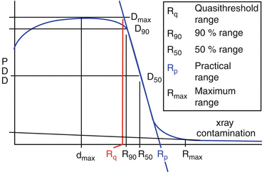
Fig. 10.2
Electron range metrics. R90 and R50 are defined by the depth of the 90 and 50 % isodose lines. A straight line is drawn between R90 and R50 and used to calculate extrapolation values. Extrapolating back to 100 % gives the Rq, while extrapolating forward to 0 % gives the Rp. Rmax is the maximum range of electrons, after which dose is entirely due to Bremsstrahlung x-rays.
Electrons: Shape of Pdd Curve
Electrons are directly ionizing so there is no charged-particle buildup like with photons.
So, why is not surface dose 100 %? (it is more like 75–95 %).
Multiple scattering causes an increase in dose at depth, so the max dose is higher than the surface dose (Fig. 10.3).
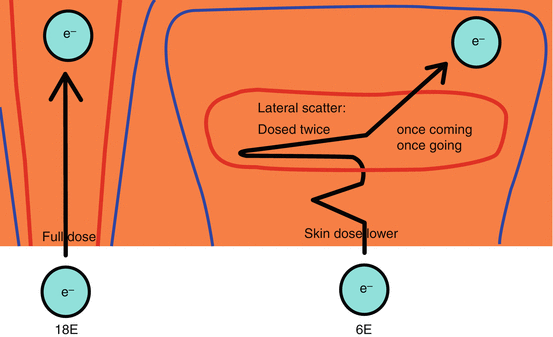
Fig. 10.3
Multiple scatter and depth dose. Scatter causes a dose buildup effect at a small depth (~1–2 cm) from the surface. Higher energy electrons scatter less, so surface dose increases with energy, unlike with photons.
After d max , dose decreases as electrons reach the end of their range (R 50 , R p , R max ).
Low energy electrons have a very sharp distal dose fall-off, while higher energy electrons have a more gradual distal fall-off (Fig. 10.4).
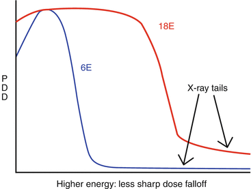
Fig. 10.4
Dose Falloff from Electrons. Higher electron energies have a longer range, but they suffer from a less sharp distal dose falloff and a larger Bremsstrahlung X-ray “tail”.
Beyond R max , dose decreases to a low but non-zero number.
Bremsstrahlung X–rays (<5 % of electron dose).
This dose increases with electron energy and with materials in the electron beam. The scattering foil is a major contributor to bremsstrahlung.
Bremsstrahlung dose is greatest at central axis and less at field edges.
Electrons: Energy Spectrum and Range
The nominal energy of an electron beam is equal to the electron energy in the accelerator. This is a mono-energetic value.
So “15 MeV electrons” have exactly 15 MeV just before passing through the window of the waveguide.
Electrons lose energy in the scattering foil, monitor chamber, and while traveling through air.
This causes “energy straggling”, resulting in a poly-energetic spectrum (Fig. 10.5).
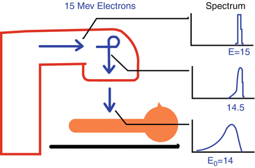
Fig. 10.5
Electron beam spectrum. Electrons exit the waveguide with a single well-defined energy, but their energy begins to “straggle” even inside the linac head. Electron energy at the patient’s surface, E0, is slightly lower than the nominal energy. Energy spectrum is shown at various locations.
E = Nominal Energy (i.e., 15 MeV).
E 0 = Mean energy at patient surface = 2.33 MeV * R 50 (by definition).
E 0 is the primary measure of beam quality for electrons.
This number is always slightly less than the accelerator energy. 15 MeV electrons may have an E 0 = 14 MeV.
Within the patient (or phantom), mean electron energy decreases linearly with depth z, as described by Harder’s equation
Stay updated, free articles. Join our Telegram channel

Full access? Get Clinical Tree


