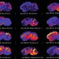
Fig. 1
Schematic representation of strategies used for microproteomics analysis using Liquid MicroJunction (LMJ) . The upper panel shows methods to define regions of interest (ROIs) according to highlighted regions, such as by histological staining, immunohistochemistry, or molecular histology profiling by Mass spectrometry Imaging . Once the ROI is defined, microproteomics can be performed in two different ways. Localized on-tissue microdigestion of fresh/frozen (fr/fr) or Formalin-fixed and paraffin-embedded (FFPE) tissue is combined with subsequent liquid microjunction to extract digested peptides . Intact proteins can also be extracted directly from ROIs of fr/fr tissue
2 Materials
2.1 Washing Steps for Fresh Frozen Tissue Preparation
- 1.
Ethanol 75% (−20 °C): 75 mL of absolute ethanol (≥99.8%) and water (HPLC grade) to 100 mL. Prepare fresh. Store at −20 °C.
- 2.
Ethanol 95% (−20 °C): 95 mL of absolute ethanol (≥99.8%) and water (HPLC grade) to 100 mL. Prepare fresh. Store at −20 °C.
- 3.
Chloroform (−20 °C): 100 mL of chloroform (≥99.9%). Store at −20 °C. Chloroform is toxic when inhaled , so work in the hood.
2.2 Dewaxing Steps for FFPE Tissue Preparation
- 1.
Xylene: 100 mL of xylene (≥99.9%). Xylene is toxic when inhaled, so work in the hood.
- 2.
Ethanol 100%. Prepare fresh.
- 3.
Ethanol 95%: 95 mL of absolute ethanol (≥99.8%) and water to 100 mL. Prepare fresh.
- 4.
Ethanol 75%: 75 mL of absolute ethanol (≥99.8%) and water to 100 mL. Prepare fresh.
- 5.
Ethanol 30%: 30 mL of absolute ethanol (≥99.8%) and water to 100 mL. Prepare fresh.
- 6.
Water: 100 mL of water (HPLC grade). Prepare fresh.
2.3 On-Tissue Digestion Using a Microspotter
- 1.
- 2.
Methanol 50%: 50 mL of absolute methanol and water to 100 mL. Prepare fresh. Methanol is toxic, so work in the hood.
- 3.
Aqueous TFA solution 0.1%. TFA is toxic, so work in the hood.
- 4.
2.4 Modification on Advion Triversa Nanomate with LESA Option
- 1.
Advion Triversa Nanomate platform (Advion BioSciences Inc., Ithaca, NY, USA), with Liquid Extraction Surface Analysis (LESA) option .
- 2.
Chipsoft software (version 8.3.1.1018 build 101018) with the Advance User Interface (AUI) enabled and LESA Points software (version 1.1.0.0).
- 3.
- 4.
Non-conductive tips, 0–10 μL.
2.5 Liquid Microextraction of Digested Peptides
- 1.
Aqueous TFA solution: 0.1% TFA is toxic, so work in the hood.
- 2.
Methanol 70%: 7 mL of absolute methanol and water to 10 mL. Prepare fresh. Methanol is toxic, so work in the hood.
- 3.
Acetonitrile 80%: 8 mL of Acetonitrile and water (HPLC grade) to 10 mL. Prepare fresh. Acetonitrile is toxic, so work in the hood.
2.6 Liquid Microextraction of Intact Proteins
- 1.
SDS Solution: Resuspend SDS in Tris–HCl (pH 8, 0.1 M) with DTT (50 mM) to obtain a final concentration of 1% (w/v).
- 2.
CHAPS Solution: Resuspend CHAPS in Tris–HCl (pH 10, 0.1 M) with DTT (50 mM) to obtain a final concentration of 4% (w/v).
2.7 Shotgun Analysis
- 1.
Urea solution: Resuspend urea in Tris–HCl (pH 8.5, 0.1 M) to obtain a final concentration of 8 M.
- 2.
2.8 Protein Identification and Quantification
- 1.
MaxQuant (latest version is available at: http://www.coxdocs.org/).
- 2.
UniProt reference protein database for the organism of interest.
2.9 Quantification -Based Mass Spectrometry Profiling
- 1.
Perseus Framework (latest version is available at: http://www.coxdocs.org/).
- 2.
TIGR Multiexperiment viewer (MEV v4.9).
3 Methods
This methodological part is decomposed according to the workflow presented in Fig. 1. We present here only methods for microdigestion and/or LMJ. MALDI MSI is not the principal aspect of this method description. Detailed protocols for tissue preparation for MALDI MSI could be found in our previously published chapter for Formalin Fixed, Paraffin embedded (FFPE) , or Fresh and frozen (fr/fr) tissue sections [23]. Complementary to data obtained from MALDI-MSI, information from histology or immunohistology could be used to determine regions of interest. Concerning MALDI-MSI, different algorithms are used to realize spatial segmentation of spectra in order to obtain molecular histology [24]. In this case, regions are defined only based on their molecular signatures and not on histological information like the morphology of cells for example. Molecularly different ROIs are selected and localized microproteomics is performed on these regions.
We primarily describe the steps leading to the preparation of tissue sections prior to microproteomics analysis, which is an extremely important part of the method. For fresh frozen tissue sections, an important first step is the washing of the sample, like in MALDI MSI . The use of an ethanol bath for fixation of proteins and removal of endogenous salts and peptides is coupled to a wash using chloroform for depletion of lipids . These washes prevent the extraction of these components as they could interfere with subsequent analysis after microdigestion. In the case of the extraction of intact proteins, washing the fresh frozen tissue section is also crucial. Indeed, LMJ using detergents , which act like a “liquid microdissection,” removed the totality of the tissue at the junction point. Complete removal of the tissue is observed in the case of the washed section, contrary to the unwashed one. Without washing steps, a severe decrease of the number of proteins identified is observed [22]. In the case of trypsin digestion (and for intact protein extraction in fact), the different washing steps destabilize the lipid bilayer membrane of the cells allowing a better penetration efficiency for the trypsin or for the detergent .
For FFPE tissue sections, in addition to the classical paraffin removal steps, an antigen retrieval step has to be performed. This step allows for better enzyme accessibility of the cleavage sites and significantly increases the efficiency of digestion. Antigen retrieval is very specific to the tissue composition and needs to be optimized and chosen wisely. Fixation using paraformaldehyde prevents extraction of intact proteins due to crosslinks formed between molecules. The only way in this case is to use a microdigestion step prior to extraction.
Concerning the parameters for microdigestion, the use of an automatic microdispenser that is accurate in terms of locating the dispense position and precise in terms of repeatability of deposition is important. A chemical inkjet printer permits this type of precision. In this case, the digested region will delimit the analysis region so that even if larger microextracted areas are achieved with LMJ, the area where it will extract digested peptides will remain defined by the microdigested region. This allows the analysis of features smaller than 300 μm using a chemical inkjet printer for trypsin deposition. The volume and number of the droplets are also important points to optimize. In the case of microdigestion for a microproteomics experiment, the digestion needs to be controlled by maintaining the droplet formed on the surface of the tissue. Figure 2a shows the droplet of solution containing trypsin formed on the surface of the tissue section. For this example, the digested region is 672 μm in diameter (Fig. 2b). If the volume of the droplet is not enough, the efficiency of digestion will be decreased but if the droplet size becomes too large, an increase of the diameter of the region will lead to a less precise analysis. These parameters are dependent on the composition of the tissue and need to be adapted for each situation. If the droplet is correctly maintained during 2 h, no additional incubation time is needed. As observed in Fig. 2b, the digested region is well defined and no spread is observed. Figure 2c shows the previously digested region after LMJ. The size of the extracted region is more than 1.5 mm in diameter (1876 μm). In this case, the size of the analysed region is defined by the size of the digested region, as shown previously [17].

Fig. 2
(a) Optical image of a microdigestion droplet maintained on the surface of a tissue section. (b) Magnified view of the digested region and (c) the same region after LMJ. The previously digested region is observed inside the extracted region of 1.8 mm in diameter
Concerning liquid extraction, the size of the microjunction is defined by several parameters, such as the volume of solution used to create the junction and the distance between the surface and the end of the collecting tip. These parameters need to be tested properly before the extraction process. Typically, a distance of dispense between 0.2 and 0.4 mm and a volume between 0.6 and 0.9 μL will lead to a junction of around 1 mm in diameter. Figure 3 presents the three possibilities for the setting of the distance between the tip and the surface to analyze. In Fig. 3a, the tip is too high and no liquid is in contact with the surface. On the other hand, in Fig. 3c, the tip is too close and the junction is not visible. In this case, extraction could occur but the amount of solution is not enough to be able to do optimal extraction. In Fig. 3b, the distance and the volume are optimal to maintain a microjunction. A repeat of the extraction cycle could be done. For the extraction of digested peptides , it has been observed that successive cycles using a combination of different solutions will increase the extraction efficiency. For intact proteins, repeating the number of cycles realized in the same position is also possible. However, too many repeats will increase the size of the junction and decrease the precision of the analysis. Three cycles of extraction are normally enough to remove all the tissue in the extracted region and if some tissue remained, an additional extraction step can be performed. Figure 4 shows three scenarios that could be observed after liquid microjunction . In Fig. 4a, total removal of the tissue is observed and this corresponds to the optimal settings. Figure 4b presents a partial removal of the tissue. Optimization is needed either by choosing a more appropriate detergent or by increasing the number of extraction cycles. If spreading of liquid is observed (Fig. 4c), the dispensed volume needs to be adjusted or the number of extraction cycles is too much.


Fig. 3
Photograph showing varying distances between the conical tip and the sample. (a) The dispensing end of the tip is too far from the surface and no junction is formed. (b) Optimal distance between the tip end and the surface with formation of a liquid microjunction (blue liquid used for a better visualization). (c) The tip end is too close to the surface and the contact between the liquid and the tissue is limited
Fig. 4
Photomicrographs of the extracted region showing a total (a) or a partial (b) removal of the tissue or spread of liquid after LMJ and (c) a partial spread of solution around the extracted region
Concerning the solution used to perform extraction after microdigestion, various solvents are employed. Combining three consecutive extractions with different solvents significantly improves the extraction. Optimizations have been done using first an acidic solution to inactivate the enzyme and extract the more hydrophilic peptides . Then, two consecutive extractions using solutions of methanol and acetonitrile were performed to extract all remaining peptides. Extraction of intact proteins is performed using a solution containing detergents like CHAPS or SDS. Solutions containing only organic solvents extract a small fraction of major proteins. Deeper analysis needs the use of a detergent solution to be sure to extract the maximum amount of proteins at the junction.
After LMJ, the extract is dispensed on a collecting tube and could be used for various analyses that can be found in previously published studies [17, 18, 22].
3.1 Preparation of Fresh/Frozen Tissue Section
- 1.
Tissue sections with a thickness of 20 μm are used (see Note 5 ) and mounted sections are stored in a sealed container at −80 °C until use.
- 2.
Before use, slides are warmed at room temperature under vacuum to prevent water condensation on the surface of the frozen slide.
- 3.
After complete drying, the glass slide is washed using different solutions. The washing step is a critical point and skipping this step could lead to a decrease of the number of proteins extracted.Related posts:
 “A Future Amalgamation Between the Scientist and the Clinician?”
 Peptide Imaging: Maximizing Peptide Yield, Optimization of the “Peptide Mass Fingerprint”
 MALDI-MS Imaging in the Study of Glomerulonephritis
 DESI Mass Spectrometry Imaging (MSI)
 Imaging MS of Rodent Ocular Tissues and the Optic Nerve
 Laser Ablation Inductively Coupled Plasma Mass Spectrometry Imaging of Plant Metabolites

Stay updated, free articles. Join our Telegram channel

Full access? Get Clinical Tree


