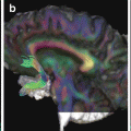
Fig. 13.1
Nonexhaustive selection of preclinical applications
Special Considerations in the Clinic
When embarking on DTI data collection in a clinical setting or in the context of clinical research, there are a number of considerations that could be taken into account that will guide the way data will be acquired and how it is analyzed. Each of these is summarized below.
Scan Population: Whom Are We Going to Scan?
Nature of Patient Group/Pathology
Different clinical groups present specific challenges. For example, acquiring DTI data on neonates is clearly different in practice to acquiring data on adults. Similarly, acquiring DTI data in the presence of substantial traumatic brain damage will present more challenges than scanning patients with more subtle organic pathology, such as in prodromal schizophrenia . These are extreme cases, however, they illustrate the importance of considering the nature of the patient group and their pathology, when acquiring and analyzing DTI data. Some specific examples are provided in the following chapters, however, more generally, you may consider the following.
Age
The brain changes significantly over the course of the lifespan, and this in turn is reflected by alterations in DTI parameters that are modulated by structural changes as the brain develops and ages. Such changes are not uniform across the brain, and throughout each stage of life fiber pathways are developing and degenerating at different rates [15, 16]. Understanding when these changes occur is useful both when considering the design of DTI research studies and when interpreting clinical findings in the context of other imaging or histopathological data.
Developmental Phase
Although the possibility of acquiring in vivo DTI data and performing tractography in utero has been demonstrated [17], the numerous challenges associated with acquiring such data limits the techniques use in clinical practice. Nevertheless, several groups have applied DTI to study fetal brain development both ex-vivo, using postmortem brains [18, 19] and in-vivo, by scanning preterm infants [20]. As DTI provides contrast in the absence of myelination, these fascinating studies have been able to build on prior histological knowledge to provide further insight into brain development. Far from being small, simplified versions of the adult brain, the fetal brain is a unique, and continuously changing with transient structures such as the ganglionic eminence [21] and fiber bundles that may disappear during fetal life, at term or in early childhood. Others bundles, such as the superior longitudinal fasiculus, may not be detectable until the third trimester or even at birth [22]. Unlike in later developmental stages where DTI is most useful in delineating white matter structure, in the fetal brain, the organization of the cerebral wall is also apparent. Notably, the presence of radially oriented structures that guide neuronal migration to the cortical surface between the second and third trimesters, give rise to anisotropy perpendicular to the cortical surface, which results in high FA in the cortex. As anisotropy decreases with increasing microstructural laminar and columnar complexity in later development, FA, in turn, becomes much lower in the cortex.
Knowledge of such variations in anatomical organization and DTI parameters during fetal life will become increasingly valuable in future clinical applications involving preterm and newborn infants [23].
Typically, as myelination progresses, anisotropy within the white matter continues to increase and diffusivity decreases rapidly during the course of brain development, with the most dramatic changes occurring in the first 2 years of life. After this period, the rate of change decreases as the brain approaches adult proportions in mid-childhood [24]. For a detailed review on the topic of white matter development during the fetal, neonate and infant stages, see Dubois et al. [25].
In order to investigate DTI changes during development, various groups have proposed a series of age-specific population atlases including data from neonates, infants, and children through to adolescence [15, 26]. Although different studies are broadly in agreement with respect to the identification of increases in anisotropy followed by decreases in diffusivity during development, the precise trajectory of such changes remains to be confidently elucidated owing to the myriad challenges associated with DTI data collection and analysis.
Senescent Phase
It is generally accepted that the age-related decline in white matter volume in late adulthood is an indicator of senescence. Precisely when this phase begins and development end is unclear, however DTI data suggests it may occur as early as the fourth decade [27]. In line with heterogenous DTI changes during development, FA and diffusivity changes during senescence are equally regionally variable. One prevailing view is that of retrogenesis, which describes how fiber tracts that are myelinated the latest, are the most vulnerable to neurodegenerative processes. Such early myelinated fibers, which reach maturation in utero or perinatally (e.g. primary motor fibers) are thought to be more robust than those maturing later (e.g. SLF). An allied phenomenum is the concept of an anterior-posterior gradient of diffusion changes, whereby age-related decline in FA is greatest in frontal and parietal WM compared to occipital WM [28].
Advancing age is associated with a loss of white matter volume, FA decrease, and increase in diffusivity measures. There are also other age-related phenomena that may impact upon DTI metrics, which deserve consideration. A common feature of elderly MRI scans are hyperintense lesions on T2-weighted fluid-attenuated inversion recovery (FLAIR) images. These “white matter hyperintensities” (WMH) reflect regional leukaraiosis arising from multiple histopathologic processes, including ischemia/infarction, demyelination, inflammation, gliosis and rarefaction, and amyloid angiopathy [29]. WMH typically occur in a regionally characteristic pattern, with focal or punctuate lesions that may become confluent, occurring in the deep and subcortical white matter; and periventricular WMH characterized by smooth, confluent bands and periventricular caps. The former are thought to arise from primarily vascular pathology, whilst the latter periventricular lesions are believed to have a non-ischaemic origin [30]. Historically, the boundaries of WMH have been defined by a sharp change in intensity on FLAIR images allowing relatively trivial qualitative clinical ratings and automated segmentation. However, a more contemporary view is that WMH reflect a late, severe stage of damage that extends beyond the hyperintense boundary; a view supported by recent work combining DTI and FLAIR to investigate the evolution of WMH [29, 31]. In this context, DTI studies in populations that include subjects with WMH , should take into account not just the clearly delineated WMH, but also consider potential broader microstructural changes and partial volume effects.
A less widely considered factor in the context of DTI, is the change in brain iron content with advancing age. Ferritin, a protein controlling the storage and release of iron, decreases in white matter and increases in subcortical gray matter [32, 33]. As iron changes MR-signal intensity and causes susceptibility artifacts, any increase in iron-content will influence DTI metrics.
Effect on Brain Structure
Different clinical groups will share both overlapping and distinct neuroanatomical features. For example, infants will share similar features to other infants, but will have markedly different brains to older typically developing children and adults. Similarly, healthy adults of comparable age will share similar gross anatomy, but adults within that age category who have multiple sclerosis , will have brains with regionally altered white matter composition. It is important to recognize and consider such differences in the context of DTI as the size, shape, and composition of the brain impacts DTI parameters in a number of ways. For example, by differentially changing the homogeneity of the magnetic field, particularly at tissue interfaces such as the frontal and temporal sinuses , there will be differential degrees of gradient susceptibility distortions and signal loss. In such regions, the measured diffusion signal will be corrupt and therefore any derived DTI metrics will be inaccurate. Although it may be possible to recover some information by applying offline postprocessing correction strategies such as informed RESTORE [34] or field-map-based techniques [35], attempting to reduce these effects during data acquisition are encouraged.
Brain geometry will also determine the extent of partial volume effects (PVE) . For example, larger brains may be expected to have larger fiber bundles and therefore less PVE-contaminated voxels relative to smaller fiber bundles. As DTI parameters represent a voxel-average scalar measure, PVE modulate DTI parameters [36]. It follows that systematic differences in PVE may introduce bias into tract-based statistics.
Almost all DTI processing will be performed in another reference space than the native image space. This is because image quality correction techniques that attempt to reduce the effect of eddy currents and motion, require the image to be transformed using image registration techniques . For example, by mapping the DWIs to the non-DWI (b0), or to an anatomical scan. Image registration is also required for any sort of group analysis where equivalent voxels/anatomy needs to be compared between subjects equitably. In simple terms, registration algorithms seek to find common features between images in order to match them. In the presence of large differences in brain structure such as gross atrophy or lesions, the registration will perform more poorly. In the case of individual datasets with atypical anatomy and/significant pathology, special attention should be paid to assessing the quality of correction strategies. In the case of group comparisons, the template space should match the study population as closely as possible (e.g. pediatric). Ideally, a population atlas derived from combining all the subjects data, should be used, or alternatively, a high quality template developed by the neuroimaging community (see Chap. 10 for further details).
Effect on Patient Mobility
The sequences used to acquire DTI data (e.g. EPI), are particularly susceptible to motion. In the absence of real-time motion correction, it is therefore preferable for the subject to remain as still as possible for the duration of the scan. There are a number of clinical groups where this may be more challenging. For example: children, patients with motor disorders or hyperkinesia due to other pathology or medication, and patients that are particularly restless, agitated, or anxious (including patients with active psychiatric symptomatology). When such subjects are included in group studies, it is particularly important to recognize the potential for bias if there are more motion artifacts in the patient data compared to the control data.
Aside from increased motion, patients may also present with reduced or restricted mobility. In such cases, patients may not be positioned optimally within the scanner, or suffer discomfort.
Degree of Patient Compliance
The patient’s motivation for having a DTI scan and their understanding of the procedure may impact on the success of data collection. A significant number of patients may refuse or terminate scanning early due to anxiety, typically arising out of claustrophobia [39]. Aside from the risk of incomplete data collection, data quality may be reduced by motion artifacts arising from gross patient movement, increased respiration and swallowing. DTI sequences involving EPI are characterized by a persistent, loud high frequency repetitive beep and strong gradients that may vibrate the scanner table. These features may startle or unnerve anxious patients. However, careful explanation and the provision of realistic expectations of the scanning process has been shown to reduce patient anxiety and increase compliance, and therefore reduce unwanted motion effects or early termination of scans [40].
DTI in Children
Aside from the analytical challenges associated with changes in brain development described above, there are other practical factors that should be considered when acquiring DTI data in children (for a detailed review, see [41]). These considerations can be broadly divided into the following themes.
Child-Centered Approach to Scanning
Acquiring DTI data on children is challenging across all age groups, as the scanning procedure requires that they lay still in an unfamiliar, noisy restrictive space for a relatively long period of time. This can be challenging enough for adults, let alone for younger children who may not comprehend what is happening and why. At best, this may result in poor quality data due to excessive bulk motion artifacts, at worst; it will result in significant distress and termination of the scan and thus incomplete data acquisition. Approaches to avoid these scenarios are therefore necessary, both for the sake of clinical care and the child’s well-being, and also to prevent potential bias due to motion artifacts in case–control group studies. The most appropriate approach will be guided by the age of the child and the context of the scanning protocol . For example, if the DTI scan is required for clinical purposes, such as for presurgical planning, where good quality data is essential to clinical care, the child will typically be sedated. If the child is voluntarily taking part in a clinical research study, a different approach will be required. Babies are encouraged to sleep and pre-school or school-aged children are typically awake.
Broadly, a child-centered approach involving parents and carers is recommended, both in the scanning environment and in communication. This could involve a decorated waiting area, the provision of child-size furniture and toys, and child-friendly uniforms for medical staff. Some institutions have a mock scanning facility, which can be used to introduce the child to the scanner by replicating the scanning experience in a safe, controlled manner. Child-appropriate scanning equipment is useful, for example, a smaller head coil, foam padding and blankets, ear-plugs/headphones to reduce scanner background noise (SBN) , and the opportunity to stream children’s music/videos within the scanner.
With regard to communication, it is important to engage both parents/carers and children, as parental anxiety and misunderstanding will translate to the child. Furthermore, parents are required to provide written informed consent in addition to verbal consent from the child, and should therefore fully comprehend the purpose and procedures surrounding the DTI scan. Some ethical committee regulations may also require written consent from children, typically over 12 years of age. Appropriate communication can take many forms but should always make use of child-specific and/lay terminology and be presented in a calm, unhurried, and friendly manner. Aside from general verbal instructions, it is useful to offer illustrated information booklets, brochures, and videos. Beyond these basic approaches , a variety of other methods can be used, for example drawing on behavioral and situational training techniques [41]. Further technical details about acquiring DTI data in children can be found in Chap. 6.
Ethical Considerations
A complete discussion of ethical considerations associated with the use of DTI data is beyond the scope of this book. However, a few points deserve mention. Firstly, the same fundamental principles apply to DTI scanning as to any other noninvasive imaging procedure. Informed consent should be obtained and the DTI examination should conform to local institutional regulatory ethical standards and those required within broader national and international frameworks e.g. International Code of Medical Ethics, Declaration of Helsinki and The Belmont Principles.
Secondly, the decision to use DTI in a clinical setting should not be undertaken without due consideration of the limitations of the technique, particularly with regard to risk-assessment. The question arises as to whether or not to use DTI data if it is available i.e. is suboptimal, potentially incorrect data about white matter anatomy better than no information at all? This is especially pertinent in the context of surgical planning. For example, it has been clearly demonstrated that DTI-based fiber tractography does not provide a complete representation of anatomy and underestimates the size and cortical extent of white matter tracts [42], therefore using the technique without due regard to this limitation is potentially harmful. In practice however, surgeons typically draw on a range of imaging and electrophysiological techniques for presurgical risk assessment and planning. They are also more likely to use the color FA maps in the first instance, with DTI-based tract reconstructions providing a coarse assessment of tract displacement (see Chap. 20). When used responsibly, this complementary information may improve surgical outcomes [43]. Surgical planning is an illustrative example, however, the decision to use DTI data in other clinical settings should be guided by the acting physicians duty of care.
Thirdly, there are different ethical issues associated with acquiring data for clinical care and for preclinical research, and due attention such be paid to this distinction. Of particular note are issues associated with managing incidental findings [44] and the difficulty patients may experience distinguishing between a diagnostic brain scan and one used for research purposes i.e. the “therapeutic misconception ” (e.g. clinical study participants may incorrectly assume the scan forms part of their medical record and will be used to identify pathology) [45]. Finally, there is an increasing trend towards data-sharing within the neuroimaging research community and a concerted effort to understand brain connectivity, often incorporating DTI data. This sharing of clinical research data for secondary analysis presents additional ethical challenges, particularly relating to informed consent (see Brakewood and Poldrack [46], for a detailed review).
Scanner Resources: What Equipment Is Available?
Hardware
Magnetic Field Strength
Clinical MR systems typically used for routine DTI scanning of humans are 1.5 T or 3 T, although a small number of specialist research centers offer the possibility of scanning (predominantly healthy) subjects at 7 T. As DTI parameters should not be dependent on the static magnetic field strength, the reproducibility of measures at different field strengths is largely dependent on signal-to-noise ratio and the effect of artifacts [47]. It is commonly accepted that scanning at higher field strengths provides increased signal-to-noise, therefore one would expect higher fields to equate with higher quality. Although this is the case for conventional imaging, the competing decreases in T2 time and increased b0 inhomogeneity associated with increasing field strength, coupled with increased distortions due to eddy currents, magnetic susceptibility, and chemical shift artifacts, off-set the gain in image quality in DTI. Nevertheless, it has been shown that the uncertainty of fitted DTI parameters decreases with increasing field strength, which may impact positively on fiber-tracking results [47]. Additionally, the use of multichannel phased-array head radio-frequency coils in place of a traditional birdcage coil allows the use of parallel imaging techniques such as sensitivity encoding (SENSE), array spatial sensitivity encoding technique (ASSET), and generalized auto-calibrating partially parallel acquisition (GRAPPA) to improve DTI data quality. At moderate acceleration factors (e.g. 2 or 3) these techniques may reduce susceptibility-induced geometric warping artifacts and T2 effects by allowing a reduced EPI echo train and TE time [48].
Gradient System
Aside from the static field, the number and strength of transmit and receive gradient coils contributes significantly to DTI data quality. Additionally, the gradient duty cycle determines how many 2D images can be acquired per TR. Modern scanners will have larger duty cycles and therefore allow more data to be collected in less time. Increasing gradient strength allows stronger diffusion weighting and read-out in a shorter period of time. In turn, this means TE can be reduced, which improves data quality by decreasing susceptibility effects. The rate at which the gradients can be turned on and off, the slew rate, is also important. Fast slew rates are preferable for DTI. However, the rapid rise and fall of strong gradients can induce image distorting eddy currents and mechanical vibrations. More significantly for the patient, rapid gradient switching can cause peripheral nerve stimulation leading to involuntary muscle contractions. For safety reasons, the maximal gradient amplitude and slew rate are therefore limited. Typical clinical systems operating within these safety limits use gradients in the order of 40–80 mT/m maximal gradient amplitude and 150–200 mT/m per millisecond maximal slew rate, although new systems with stronger gradients and faster slew rates are emerging on the market and in specialist research centers.
Peripheral Equipment
DTI data quality may be improved by using nonstandard equipment provided by the scanning manufacturer or developed by and for researchers. For example, using multi-channel phased array coils instead of birdcage coils to boost SNR and to allow parallel imaging a simple way to improve DTI data quality. However, it should be noted that increasing the number of component coils may introduce a bias field which favors cortical coverage over deep white matter and therefore may be less optimal for DTI in these regions. Peripheral cardiac-gating is commonly supplied by MRI vendors and enables DTI data to be acquired only during the diastole phase of the cardiac cycle, thereby reducing pulsation artifacts. At the other end of the spectrum, prospective motion correction using real-time tracking with external devices such as cameras is gaining ground in high-field research centers [49]. When such advanced equipment is available, the choice remains as to whether or not it would be appropriate to use it. For example, cardiac-gating increases scan time and may not be useful in restless subjects or when data must be acquired quickly.
Number of Scanners
This will determine to a great extent, the relative availability of DTI scanning time. Consider, how many people are using the scanner/s and how frequently. In a primary clinical setting, consider the patient turnover of the department. DTI sequences can vary in length from a few minutes to half an hour or more depending on the amount of data acquired and number of gradient directions sampled. In a small clinic with only one scanner and a high patient turnover, it is clearly more sensible to consider short sequences, and the possibility of performing long, complex longitudinal data collection may be limited. The need for standardization of protocols and effective quality assurance procedures is particularly important within a center with multiple scanners.
Software
Data Management
An often-overlooked issue when managing DTI data is how it will be stored and transferred from the scanner to other platforms. Typical DTI datasets are much larger than standard clinical scans and therefore present additional storage issues, greater burden on PACS (Picture Archiving and Communication System) and extended transfer time.
Data Storage
It is beyond the scope of this chapter to discuss DTI data storage options in detail; however, those working with DTI data should consider how and in what form it will be stored. For example, will the data be archived on PACS? Or stored off-line on a secure server? Or within a cloud computing framework? Will there be a back-up? Who will have access to this data? How will it be accessed? Who will manage the data? Will the raw DICOM data be stored, or also processed data? These are examples of issues common to all types of imaging data. The difference with DTI data however is that DTI datasets are larger and thus present a more disproportionate storage burden than other data.
Related posts:


Stay updated, free articles. Join our Telegram channel

Full access? Get Clinical Tree


