Duodenal Injury
Kavya E. Reddy
Ellie R. Lee
CLINICAL HISTORY
24-year-old male with right upper quadrant abdominal pain.
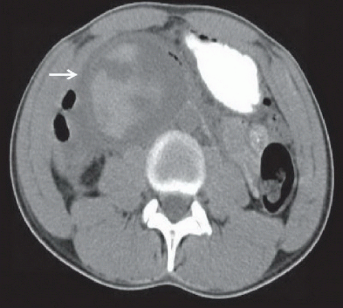 FIGURE 39A |
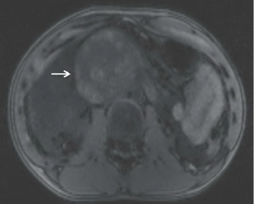 FIGURE 39B |
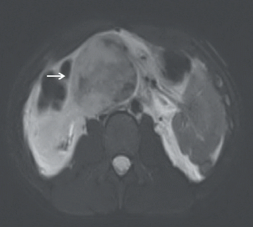 FIGURE 39C |
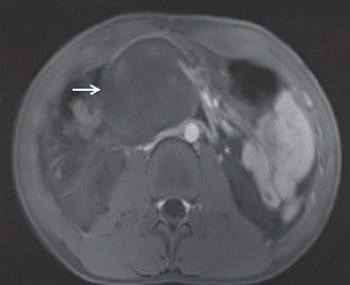 FIGURE 39D |
FINDINGS
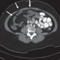
Figure 39A: Axial noncontrast CT image of the upper abdomen demonstrates a large, heterogeneous intraluminal mass in the second and third portions of the duodenum, compatible with a hematoma (arrow). There are regions of internal hyperdensity, indicating acute hemorrhage. Figure 39B: Axial T1-weighted MRI image demonstrates a large, heterogeneous intraluminal mass in the second and third portions of the duodenum showing regions of T1 hyperintensity (arrow). Figure 39C: Axial T2-weighted MRI image demonstrates a large, heterogeneous duodenal mass with regions of T2 hyperintensity, indicating hematoma (arrow). Figure 39D: Axial postcontrast T1 MRI image demonstrates no enhancement of the duodenal mass, indicating a hematoma (arrow).
Related posts:


Stay updated, free articles. Join our Telegram channel

Full access? Get Clinical Tree


