Duodenal Perforation
Shaun R. Rybak
CLINICAL HISTORY
80-year-old female with acute diffuse abdominal pain and bloating.
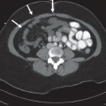
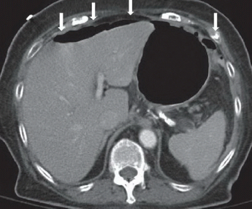 FIGURE 11A |
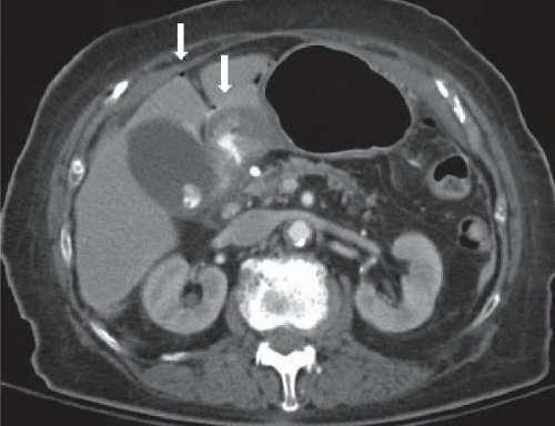 FIGURE 11B |
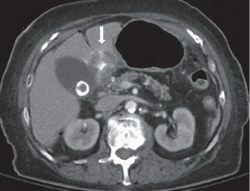 FIGURE 11C |
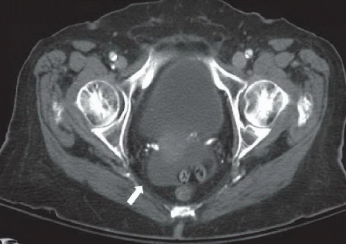 FIGURE 11D |
FINDINGS
Figure 11A: Axial contrast-enhanced CT image of the upper abdomen demonstrates pneumoperitoneum along the anterior abdomen (arrows). Figure 11B: Axial contrast-enhanced CT image of the upper abdomen demonstrates circumferential thickening of the duodenal bulb containing oral contrast. There is adjacent hyperdense fluid and a few tiny extraluminal air bubbles anterior to the duodenal bulb (arrows). Figure 11C: Axial contrast-enhanced CT image of the upper abdomen demonstrates a linear hyperdense tract extending from the lumen of the duodenal bulb through the wall, indicating oral contrast extravasation (arrow). Figure 11D: Axial contrast-enhanced CT of the pelvis demonstrates free fluid in the pelvic cul-de-sac (arrow).
Related posts:

Stay updated, free articles. Join our Telegram channel

Full access? Get Clinical Tree


