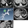
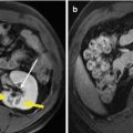
Fig. 31.1
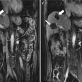
Images in a 53-year-old female with a renal clear cell carcinoma (RCC) (4.8 cm × 4.5 cm) in the right kidney treated with microwave ablation (MWA). (a) Pre-ablation contrast-enhanced ultrasound (CEUS) shows a heterogeneous hyper-enhancement lesion (arrow) in cortical phase. (b) Magnetic resonance imaging (MRI) shows a heterogeneous hyper-intense lesion in renal parenchyma (arrow) in arterial phase. (c) One month after ablation, CEUS shows the ablation zone no-enhancement (arrow) in cortical phase. (d) MR imaging showed hypo-intensity in the ablation zone (arrow) in arterial phase one month after ablation

Fig. 31.2
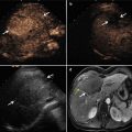
Images in a 81-year-old female with a RCC (3.6 cm × 3.4 cm) in the left kidney treated with MWA. (a) Pre-ablation US scan shows one hypo-echo parenchymal lesion (arrow). (b) CEUS scan shows one heterogeneous hyper-enhancement lesion (arrow) in cortical phase. (c) Three days after ablation, CEUS shows a very small hyper-enhancement lesion adjacent to the ablation zone (arrow) in cortical phase. (d) MR imaging shows a hyper-intense lesion at the same location on T2 image, considered as residual tumor (red arrow), (e) then no-enhancement on CEUS (arrow) in cortical phase after another ablation. (f) Hypo-intense on MRI after another ablation (arrow) on T2 image
During the median follow-up period of 23 months (range 3–90 months), 93.6 % (102/109) lesions showed completed ablation (Fig. 31.3). Six of seven recurred lesions were found by both CEUS and CT/MRI (6/109, 5.5 %). The sensitivity, specificity, accuracy, positive predictive value, and negative predictive value of recurred tumor detection by CEUS were 85.7, 99.0, 98.2, 85.7, and 99.0 %, respectively. Our results indicate that CEUS may be an effective alternative to CT/MRI in the follow-up of RCC after percutaneous MWA, which is similar to the results after RFA reported by Maria Franca Meloni in 2008 [17].


Fig. 31.3
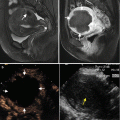
Images in a 73-year-old female with a RCC (3.4 cm × 3.4 cm) in the left kidney treated with MWA. (a) Pre-ablation contrast-enhanced CEUS shows a hyper-enhancement in cortical phase. (b) MR imaging showed one hyper-intense lesion exophyticly in the inferior pole of the kidney (arrow) on T2 image. (c) Three days after ablation, CEUS showed the whole lesion no-enhancement continuously in cortical phase. Computed tomography (CT) imaging showed one hypo-intense lesion in arterial phase 1 month (d), 3 months (e), and 12 months (f) after ablation, and the lesion shrunk gradually obviously (arrow)
Meanwhile several studies reported that CEUS played a promising role in evaluating the short-term and long-term therapeutic effects of RCC following different ablations. As shown in Tables 31.1 and 31.2, CEUS is an effective method in assessing the therapeutic effect during the follow-up of RCC followed with RFA, MWA, and cryoablation [3, 4, 17, 23, 24]. Just as conventional US, CEUS is subject to unclear lesion display for abdominal gas shielding or relative operator dependence of US. Sometimes it could be avoided by changing the posture of patient and cleaning the bowel. Otherwise, this situation may affect the assessment of the therapeutic efficiency after ablation.
Table 31.1
The CEUS evaluation performance on assessment of the ablation effect for RCCs in short-term (<3 months) follow-up
Author | Num | Ablation | Follow-up (days) | Sensitivity (%) | Specificity (%) | Accuracy (%) | PPV (%) | NPV (%) |
|---|---|---|---|---|---|---|---|---|
Hoeffel et al. [3] | 66 | RFA | 1 | 64 | 98 | N/A | 82 | 92 |
Hoeffel et al. [3] | 66 | RFA | 42 | 79 | 100 | N/A | 100 | 95 |
Li et al. [24] | 83 | MWA | 3 | 100 | 97.9 | 98.2 | 86.7 | 100 |
Barwari et al. [23] | 45 | Cryoablation | 90 | NA | 92 | N/A | N/A | 77 |
Table 31.2
The CEUS evaluation performance on assessment of the ablation effect for RCCs in long-term (>3 months) follow-up
Author | Num | Ablation | Follow-up (months) | Sensitivity (%) | Specificity (%) | Accuracy (%) | PPV (%) | NPV (%) |
|---|---|---|---|---|---|---|---|---|
Meloni et al. [17]
Stay updated, free articles. Join our Telegram channel
Full access? Get Clinical Tree
 Get Clinical Tree app for offline access

|
