Elbow Joint
Cubital Angle (Carrying Angle of the Elbow)
Various techniques have been described for measuring the cubital angle (synonym: carrying angle) of the elbow joint. The humerus–elbow–wrist angle of Oppenheim, which is somewhat more accurate than the humeroulnar angle, has become well established in the planning of corrective osteotomies and as a follow-up parameter in clinical studies. The Baumann angle can be used in children to make an approximate, indirect measurement of the joint axis in cases where the joint cannot be examined in full extension because of injury or immobilization.

Humerus–Elbow–Wrist Angle of Oppenheim
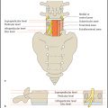
The humerus–elbow–wrist (HEW) angle of Oppenheim ( Fig. 6.1 ) is determined on an anteroposterior (AP) radiograph of the arm with full elbow extension (180°) and supination. The radiograph should cover the arm from the proximal humerus down to the wrist.
The angle is measured between the humeral shaft axis and the forearm axis. The humeral shaft axis is defined by the midpoints of two lines perpendicular to the shaft spaced as far apart as possible. The forearm axis is defined by the midpoints of two lines perpendicular to the forearm bones (lateral border of the radius and medial border of the ulna).
Humerus–elbow–wrist angle of Oppenheim
• Mean value: | 10° |
• Cubitus varus: | < 5° |
• Cubitus valgus: | > 15° |
Because the cubital angle is highly variable, a radiograph of the healthy side should always be obtained for comparison when planning a corrective osteotomy. If the affected elbow cannot be fully extended, the radiograph should be taken in maximum possible extension and the comparison view taken with the healthy limb in the same position.
Humeroulnar Angle
The humeroulnar angle ( Fig. 6.2 ) is measured on the AP radiograph of the arm or elbow joint. As the ulna often has some degree of curvature, especially in children, the accuracy of the measurement can be increased by displaying the entire ulna and entire humerus on one radiograph. The radiograph should be taken with full elbow extension (180°) and supination.
The humeroulnar angle is measured between the axes of the humeral and ulnar shafts. Ideally, both axes are drawn through the midpoints of two lines perpendicular to the shaft that are spaced as far apart as possible.

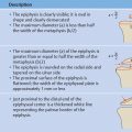
If the radiograph displays only the bone segments close to the joint, the humeral axis can be defined by reference points placed at the level where the metaphysis begins to flare and at the distal diaphysis. The ulnar shaft axis is defined by two proximal reference points at the level of the proximal ulnar border and the radial tuberosity.
Humeroulnar angle
• Mean value in females (after Keats): | 13° |
• Mean value in males (after Keats): | 11° |
Related posts:

Stay updated, free articles. Join our Telegram channel

Full access? Get Clinical Tree


