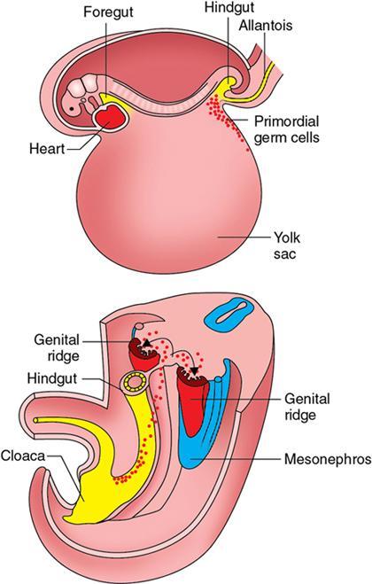
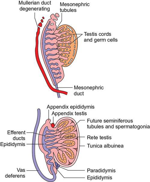
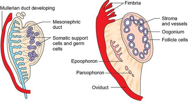
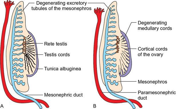
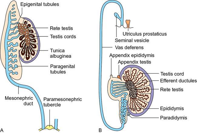
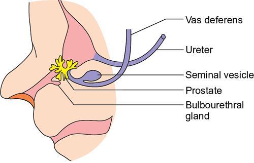
Fouzal Sex differentiation is a complex process that begins at the time of fertilization with genetic sex determination. The sexual genotype (46, XX or 46, XY) directs the development of gonads (ovaries or testis), reproductive tracts and external genitalia. Many genes including some autosomal ones are involved in this complex process. The principal gene is the SRY (sex-determining region on Y) gene in the short arm of Y chromosome (Yp11). The transcription factor encoded by this gene, SRY protein, is the testis-determining factor that initiates the male development. If the factor is absent or defective, female development is established. Thus, femaleness is a passive (i.e. default) developmental process. In both sexes, the gonads remain indifferent until 7th week of development. The formation and differentiation of gonads begin with the arrival of primordial germ cells in the intermediate mesoderm. The primordial germ cells originate in the epiblast, migrate through the primitive streak and reside among endodermal cells in the wall of the yolk sac close to the allantois during the 3rd week. During the 4th week, they migrate by ameboid movement via the dorsal mesentery of hindgut and reach the mesenchyme of the posterior body wall near the 10th thoracic level during the 5th week. The adjacent coelomic epithelium, located just medial and ventral to the developing mesonephric kidney, proliferates and thickens in response to the arrival of primordial germ cells and forms the pair of genital or gonadal ridges (Fig. 11.6.1). The primordial germ cells invade the genital ridges during the 6th week. If these cells fail to reach the ridges, the gonads do not develop. During the 6th week, cells from the coelomic epithelium form aggregates of somatic support cells that completely invest the germ cells and prevent them from degenerating. The fate of these somatic support cells in males and females are determined after 6th week. Primitive sex cords are irregular cord-like structures formed by the proliferation of the epithelium of genital ridge which then penetrate the underlying mesenchyme followed by the arrival of primordial germ cells (Fig. 11.6.2). In a genetically male embryo, the primordial germ cells carry XY sex chromosome complex. The SRY gene on the Y chromosome influences the expression of SRY protein within the somatic support cells of the gonad. In response to SRY protein, the somatic support cells begin to differentiate into Sertoli cells and envelop the germ cells. Signals arising from Sertoli cells recruit mesenchymal cells into the gonadal ridge that differentiate into interstitial cells of Leydig. During the 7th week, the differentiating Sertoli cells in the primitive sex cords, together with interstitial cells of the gonad, penetrate deep into the medulla and organize to form testis or medullary cords (Fig. 11.6.3), enclosing germ cells in the center of these cords. Towards the hilum, the cords break up into a network of thin-walled tubules of the rete testis. With development, the testes begin to round up reducing their area of contact with mesonephros and an intervening dense layer of fibrous connective tissue called the tunica albuginea separates the testis cords from the coelomic epithelium. At puberty, the testis cords canalize to form seminiferous tubules and join the rete testis tubules, which in turn connects with mesonephric ducts. The mesonephric ducts later develop into the epididymis, spermatic ducts or vasa deferentia and seminal vesicles in male. By the 8th week of gestation, interstitial cells of Leydig begin production of testosterone and induces the sexual differentiation of the genital ducts and external genitalia into male; Sertoli cells secrete anti-Mullerian hormone (AMH) that induces degeneration of the Mullerian ducts. In a genetically female embryo with XX sex chromosome complex and no Y chromosome, the somatic support cells differentiate into follicle cells (or granulosa cells) that surround individual oocytes and form primordial follicles within the ovary (Fig. 11.6.4). These follicles localize to the cortical region of the ovary. The primitive sex cords dissociate into irregular cell clusters that occupy the medullary part of the ovary and later, degenerate. They are replaced by a vascular stroma that forms the ovarian medulla and contains the stromal thecal cells that have steroidogenesis activity. Initially, both male and female embryos have two pairs of genital ducts: mesonephric (Wolffian) and paramesonephric (Mullerian) ducts (Fig. 11.6.5). The mesonephric ducts are derived from intermediate mesoderm on either side of the dorsal body wall from upper thoracic to upper lumbar (L3) segments, and grow caudally to open into the posterior wall of the primitive urogenital sinus on either side of the Mullerian tubercle. The epithelium on the anterolateral surface of the urogenital ridge invaginates longitudinally to form the paramesonephric ducts. The cranial ends of the ducts open into the abdominal cavity through funnel-shaped structures. The paramesonephric duct lies lateral to the mesonephric ducts. Then it crosses the mesonephric duct ventrally to come medial to it at the caudal end. Caudally the ducts on either side come close to each other and the caudal ends fuse to form the uterovaginal canal. The caudal tip of combined ducts forms a small swelling called the Mullerian or paramesonephric tubercle which projects into the posterior wall of the urogenital sinus. Between 8 and 12 weeks, the initial secretion of testosterone stimulates the differentiation of the mesonephric ducts. Major part of the mesonephric duct obtains a thick muscular coat and differentiates into the spermatic cord or vas deferens (Fig. 11.6.6). The cranial most end of mesonephric duct degenerates. The remnant left behind is called the appendix of epididymis. By 9th week, few mesonephric tubules in the region of epididymis, the epigenital mesonephric tubules, contact with the cords of future rete testis and is complete by 3rd month. These epigenital mesonephric tubules are then termed the efferent ductules, connecting the seminiferous tubules and rete testis with the epididymis. The mesonephric tubules at the caudal pole of developing testis, the paragenital mesonephric tubules, degenerate and the vestiges are termed the paradidymis. The accessory glands of the male genital system – seminal vesicle, prostate and bulbourethral gland – develop near the junction between mesonephric ducts and pelvic urethra (Fig. 11.6.7). By the 10th week, seminal vesicles sprout from the mesonephric ducts near their attachment with pelvic urethra. The mesonephric duct between the seminal vesicle and the urethra becomes the ejaculatory duct.
11.6: Embryology
Introduction
Gonads

Testis
Ovary
Genital ducts
Genital ducts in male
Related posts:


Stay updated, free articles. Join our Telegram channel

Full access? Get Clinical Tree


