Ependymal Tumors
Overview
Ependymal tumors of the CNS arise from the ependymal cells that line the ventricles of the brain or the central canal of the spinal cord and can affect any age group, although they are more common in children. Approximately 60% of ependymal tumors occur in the cerebral hemispheres, 30% in the posterior fossa, and 10% in the spinal canal. The two slow-growing grade I tumors are subependymoma and myxopapillary ependymoma, whereas more aggressive-grade tumors are ependymoma (grade II) and anaplastic ependymoma (grade III). Anaplastic ependymomas are more common in posterior fossa in children and supratentorial brain in adults. There are three rare distinct histological variants—papillary, clear cell, and tanycytic ependymomas. A recently defined distinct molecular subtype of ependymoma, the RELA fusion-positive, is an aggressive variant (grade II or III) of ependymoma occurring in the supratentorial brain in children and carries a worse prognosis. Although historically considered a single entity, ependymomas from each of the three anatomic compartments (supratentorial, posterior fossa, spinal canal) are considered biologically and genetically distinct entities.
|
Subependymoma
Definition: Subependymoma is a benign, slow-growing, often incidentally detected, intraventricular glial neoplasm arising from the subependymal glia and represents a lower-grade counterpart to ependymoma.
Epidemiology: Due to its rarity and asymptomatic clinical course, the true incidence of subependymoma is difficult to determine. It affects patients of all ages but is more common in middle-aged and older individuals.
Affected age group: The tumor usually affects middle-aged to older adults, with a mean age group of 55 to 64 years old, and without gender predilection.
Molecular and genetic profile: Chromosome 6 copy number alterations are only seen in lateral ventricule, posterior fossa, and spinal subependymomas and never in supratentorial subependymomas. Familial occurrence is well documented, but the underlying specific genetic susceptibility remains unknown.
Clinical features and standard therapy: Gross total resection of tumor leads to cure, although recurrence can be seen in rare cases.
Imaging
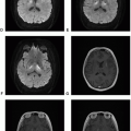
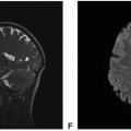
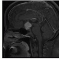
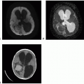
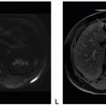
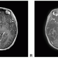
 Figure 14.3. Imaging of subependymoma of the lateral ventricle. A. Unenhanced axial CT: Right intraventricular mass with focal calcification causing obstructive hydrocephalus. B. Enhanced axial CT: Mild enhancement within the mass. C. Axial FLAIR: Hyperintense mass. D. Axial DWI: Mildly reduced diffusion. E. Axial T1-postcontrast: No enhancement within the mass. F. Axial T1-postcontrast: Irregular rim enhancement within the mass.
Stay updated, free articles. Join our Telegram channel
Full access? Get Clinical Tree
 Get Clinical Tree app for offline access

|


