Esophageal Metastases and Lymphoma
Michael P. Federle, MD, FACR
R. Brooke Jeffrey, MD
Key Facts
Terminology
Lymphoma: Malignant tumor of lymphocytes
Imaging
Ulcerated/polypoid mass of gastric cardia extending into distal esophagus
Top Differential Diagnoses
Intramural benign esophageal tumor
Esophageal carcinoma
Esophageal varices
Clinical Issues
Most common signs/symptoms
Dysphagia, weight loss, hematemesis, or asymptomatic
Esophageal metastases
Direct, lymphatic, or hematogenous spread
Direct invasion most common: Stomach carcinoma accounts for 50% of cases
Chemotherapy, surgical resection of complicating lesions (obstruction, upper GI bleed)
Complications
GI bleeding, perforation, obstruction
Prognosis
Usually poor
Treatment
Chemotherapy
Surgical resection of complicating lesions (obstruction, upper GI bleed)
Diagnostic Checklist
Check for history of primary cancer; biopsy required
Overlapping radiographic features of esophageal metastases, lymphoma, and primary carcinoma
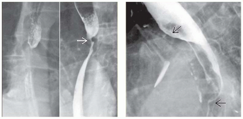 (Left) Esophagram in a woman with known lung cancer and dysphagia shows a tight stricture  of the mid-esophagus at the level of the carina. The mucosa is intact, and tapered superior and inferior margins indicate extrinsic or intramural involvement rather than a primary process. (Right) Esophagram in a man with known lung cancer and dysphagia shows a widened mediastinum and a mass in the left hilum. Note the broad shelf-like indentation of the mid-esophagus at the level of the carina. The mucosa is intact, and tapered superior and inferior margins indicate extrinsic or intramural involvement rather than a primary process. (Right) Esophagram in a man with known lung cancer and dysphagia shows a widened mediastinum and a mass in the left hilum. Note the broad shelf-like indentation  along the anterior wall of the mid-esophagus. along the anterior wall of the mid-esophagus. |
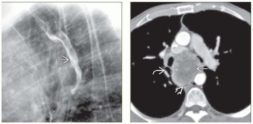 (Left) Esophagram in a 70-year-old man with a history of known bladder carcinoma, now presenting with dysphagia, illustrates extrinsic or intramural involvement of the esophagus with eccentric narrowing of the lumen
Get Clinical Tree app for offline access
Related posts:Stay updated, free articles. Join our Telegram channel
Full access? Get Clinical Tree


|
