Description
Needles are usually characterised by their mechanism of action into aspiration and core biopsy needles.
Aspiration needles
Purpose
To safely provide a sample of a few cells for cytological, biochemical and microbiological analysis. Cytology can be enough to confirm malignancy but doesn’t give any “architectural” information.
Description
Most aspiration samples are taken with standard 21G needles or spinal/ Chiba needles.
Measurements
Length (cm)
Gauge:
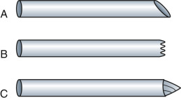
from 20-25G Most aspiration samples are taken with standard 21G needles or spinal/ Chiba needles. Simply choose a suitable size and length to reach your target. There are more specialised cutting needles available for aspiration that probably appear more often in textbooks than departmental shelves but are certainly worth considering if an initial aspiration gives scant cellular tissue ( Fig. 9.1 ).
Know how to use it
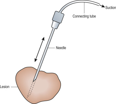
The needle is placed into the lesion under imaging guidance and then attached to a 20-mL syringe. Draw a full vacuum on the syringe while the needle is passed back and forward through the lesion ( Fig. 9.2 ). If you have an assistant, use a connecting tube or a 21G butterfly needle and get the assistant to aspirate the syringe while you manipulate the needle under guidance.
Maintain gentle suction whilst withdrawing the needle, then remove the syringe. Vigorous suction results in the tiny sample being lost in the syringe; without suction, the sample stays in the patient.
Place the sampling needle onto a slide and gently eject the needle contents onto the slide with an air-filled syringe. Also put any fluid in the sampling syringe onto a slide. Specimens may be simply air-dried but specimen preparation varies from centre to centre; check the preference of your cytologist. Specimens only contain a few cells and therefore it is best to make at least two passes.
Cutting biopsy needles
Purpose
Obtains a larger specimen for histological analysis.
Description
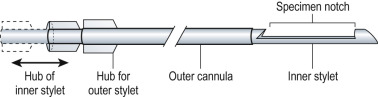
The cutting needle consists of two parts: an outer cutting shaft and an inner stylet ( Fig. 9.3 ). There are a variety of devices but most centres use automated devices, which rely on a spring loaded mechanism to automatically fire the outer cutting needle at the push of a button. Manual biopsy needles operate in the same way but are less reliable and slower. Remember for some of the designs, the needle advances when fired to take the specimen from 1-2cm beyond the firing position. The needles are always single use but the mechanism may be disposable or reusable. It is very important to understand if you have a static or forward throw device in your hand.