Esthesioneuroblastoma
Michelle A. Michel, MD
Key Facts
Terminology
Malignant neuroectodermal tumor arising from olfactory mucosa in superior nasal cavity
Imaging
Enhanced MR with bone-only CT best maps ENB for en bloc craniofacial surgery
Dumbbell-shaped mass with “waist” at level of cribriform plate
Bone CT: Bone remodeling mixed with bone destruction, especially of cribriform plate
CECT/T1 C+ MR: Homogeneously enhancing mass
Cysts at intracranial tumor-brain margin
Top Differential Diagnoses
Sinonasal squamous cell carcinoma
Sinonasal adenocarcinoma
Sinonasal non-Hodgkin lymphoma
Sinonasal undifferentiated carcinoma
Pathology
No etiologic basis or risk factors elucidated
Kadish staging system
Good predictor of outcome
Staging criteria: Kadish classification; good predictor of outcome
Histologic grading: Hyams system
Clinical Issues
Adolescent or middle-aged patient with unilateral nasal obstruction & mild epistaxis
Bimodal distribution in 2nd & 6th decades
Combined surgical resection & radiotherapy is treatment of choice
Excellent prognosis vs. other sinonasal malignancies
5-year survival rates: 75-77% overall
Recurrences in ˜ 30%
Metastases in 10-30% of patients
 (Left) Coronal graphic shows the classic features of esthesioneuroblastoma (ENB) centered below the cribriform plate and extending into the anterior cranial fossa and right orbit  . Cyst formation . Cyst formation  is noted at the tumor-brain interface. (Right) Coronal bone CT shows an ENB filling the upper nasal cavity and ethmoid sinuses. The lesion extends through the anterior skull base is noted at the tumor-brain interface. (Right) Coronal bone CT shows an ENB filling the upper nasal cavity and ethmoid sinuses. The lesion extends through the anterior skull base  . The lamina papyracea on the right is thinned and laterally displaced . The lamina papyracea on the right is thinned and laterally displaced  . . |
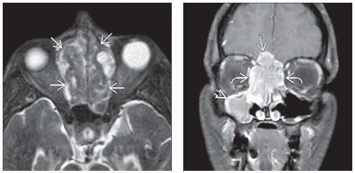 (Left) Axial STIR MR demonstrates a large, heterogeneous ENB  centered in the midline below the skull base and occupying the nasal cavity and ethmoid sinuses. The mass is predominantly hypointense in this case and causes hypertelorism. (Right) Coronal T1WI C+ FS MR shows an avidly enhancing ENB with extension into anterior cranial fossa centered in the midline below the skull base and occupying the nasal cavity and ethmoid sinuses. The mass is predominantly hypointense in this case and causes hypertelorism. (Right) Coronal T1WI C+ FS MR shows an avidly enhancing ENB with extension into anterior cranial fossa  and both orbits and both orbits  . Avid enhancement is characteristic of this highly vascular neoplasm. Note the trapped maxillary secretions . Avid enhancement is characteristic of this highly vascular neoplasm. Note the trapped maxillary secretions  . . |
TERMINOLOGY
Abbreviations
Esthesioneuroblastoma (ENB)
Synonyms
Olfactory neuroblastoma, pleomorphic olfactory neuroblastoma
Definitions
Rare malignant neuroectodermal tumor that arises in nasal cavity
IMAGING
General Features
Best diagnostic clue
Dumbbell-shaped mass with upper portion in anterior cranial fossa, lower portion in upper nasal cavity, & “waist” at level of cribriform plate
Peripheral tumor cysts at intracranial tumor-brain margin is highly suggestive of diagnosis of ENB
Location
Superior nasal cavity at cribriform plate
Smaller ENB: Unilateral nasal mass centered on superior nasal wall; local spread in nose & sinuses
Large ENB: Tumor in anterior cranial fossa with parenchymal & dural infiltration, extension into orbits
Size
Range from < 1 cm nodule to mass filling entire nasal cavity & lower anterior cranial fossa
Morphology
Polypoid mass when small; dumbbell-shaped when large
CT Findings
NECT
Bone CT
Bone remodeling causing enlargement of nasal cavity mixed with bone destruction, especially of cribriform plate area
Speckled pattern of calcification within tumor matrix unusual
CECT
Homogeneously enhancing mass
When large, may see nonenhancing areas of necrosis
MR Findings
T1WIRelated posts:

Stay updated, free articles. Join our Telegram channel

Full access? Get Clinical Tree


