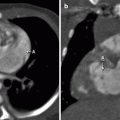
Fig. 10.1
Coronal (a) and axial (b) images from a cardiac CTA scan showing the spleen (D) and stomach (C) on the left, with the apex of the heart (A) on the left and the right atrium (B) on the right. The liver (E) is on the right. These findings are consistent with situs solitus
Polysplenia Syndrome–Type Heterotaxy
Patients with polysplenia syndrome–type heterotaxy have polysplenia; anomalous pulmonary venous return, which often is partial instead of complete; bilateral superior vena cava (SVC); left isomerism of the atrial appendages; bilateral hyparterial bronchi with bilateral bilobed lungs; and azygous continuation of the IVC.
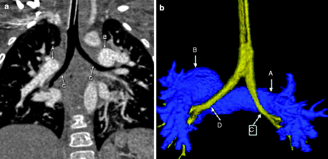
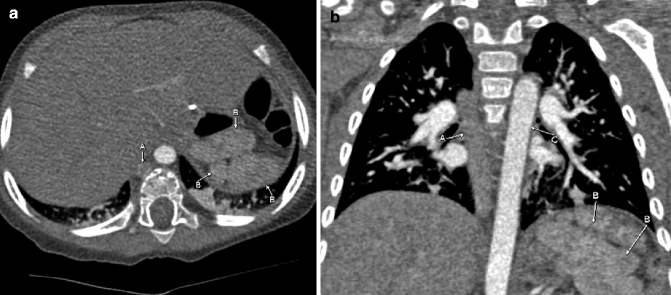
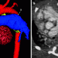
Fig. 10.2
Enhanced coronal (a) and posterior projection color-coded three-dimensional (3D; b) images showing that the right (C) and left (D) bronchi are inferior to the right (A) and left (B) pulmonary arteries, respectively. These findings are compatible with bilateral left-sided tracheobronchial branching (bilateral hyparterial bronchi) in a patient with a polysplenia heterotaxy. Notice that both bronchi (C and D) are elongated, consistent with left-sided bronchial morphology
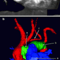
Fig. 10.3


Axial (a) and coronal (b) images from a cardiac CTA scan showing multiple spleens (B) with an enlarged azygos vein (A) in a patient with polysplenia-type heterotaxy. Notice that the descending aorta (C) is to the left of the enlarged azygos vein
Related posts:

Stay updated, free articles. Join our Telegram channel
Full access? Get Clinical Tree
