Fig. 5.1
Frontal projection three-dimensional (3D) model (a) and coronal maximum-intensity projection (MIP) image (b) from a cardiac CTA shows normal anatomy of the normal right-sided SVC (RSVC) draining the left brachiocephalic vein (LBCV) and right brachiocephalic vein (RBCV) into the right atrium (RA). Note the typical triangular shape of the right atrial appendage (RAA). LIJV left internal jugular vein, LSCV left subclavian vein, LSVC left SVC, RIJV right internal jugular vein, RSCV right subclavian vein, IVC inferior vena cava, SVC superior vena cava
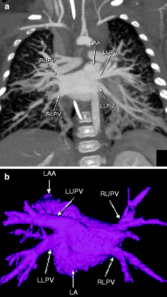
Fig. 5.2
Coronal MIP (a) and posterior projection 3D color-coded (b) images from a cardiac CTA scan demonstrating normal anatomy, with right and left upper and lower pulmonary veins (RUPV right upper pulmonary vein, LUPV left upper pulmonary vein, RLPV right lower pulmonary vein, LLPV left lower pulmonary vein, respectively) draining to the left atrium (LA) and a normal-appearing left atrial appendage (LAA). Note the elongated, tongue-like appearance of the left atrial appendage
Left Superior Vena Cava
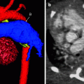
Left SVC is a common variant, especially in patients with congenital heart disease. It is important to make this finding for presurgical planning in patients undergoing SVC shunts so that there is no collateral flow around the shunt. In most instances, there is duplicated SVC physiology, with the left SVC draining blood from the left internal jugular and left subclavian veins and the right-sided systemic veins draining to a right SVC. In patients with atrial inversion that may be associated with heterotaxy syndrome, the left SVC may be the morphologic right SVC.
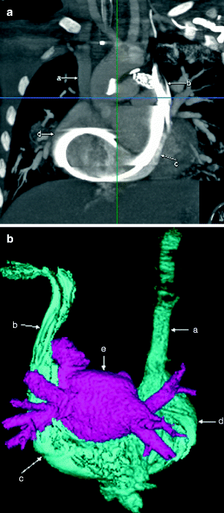
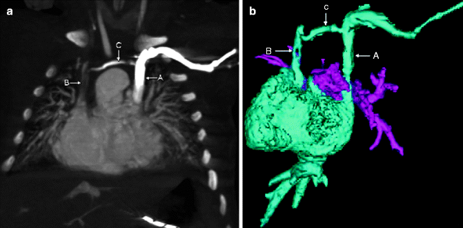
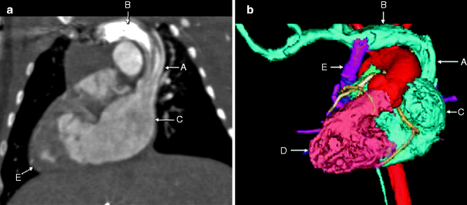
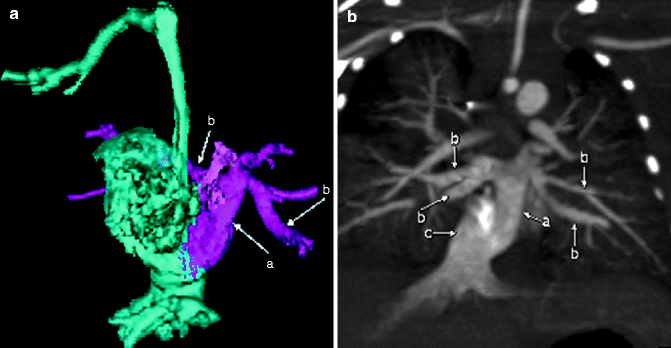
Fig. 5.3
Coronal MIP (a) and posterior projection color-coded 3D (b) images from a cardiac CT scan showing a left SVC (b) draining to the right atrium (d) via a large coronary sinus (c). Notice the distal right SVC (a) draining normally into the right atrium (d). Notice the elongated left atrial appendage adjacent to the left SVC (b)
Fig. 5.4
Coronal MIP (a) and frontal projection color-coded 3D (b) images from a cardiac CT scan showing a right (B) and left (A) SVC with a connecting brachiocephalic vein (C).
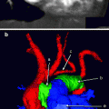
Fig. 5.5
Coronal MIP (a) and frontal projection color-coded 3D (b) images from a cardiac CT scan showing a right brachiocephalic vein (B) emptying into a left SVC (A) draining to the left atrium (C) in a patient with situs inversus. This may be associated with other congenital anomalies, such as total anomalous pulmonary venous return (TAPVR). In this case, a right-sided vertical vein (E in b) drains blood from the pulmonary veins (pink) to the brachiocephalic vein (B). Also notice that the apex of the heart is on the right, consistent with dextrocardia and known situs ambiguous in a patient with heterotaxy syndrome
Partial Anomalous Pulmonary Venous Return
Partial anomalous pulmonary venous return (PAPVR) has the following characteristics:
1.
One or more, but not all, pulmonary veins flow to a systemic vein
2.
One pulmonary lobe typically is involved, although the whole lung may be involved
3.
It typically is asymptomatic
4.
If symptoms occur, they usually are the result of right heart overload from increased volume
5.
It is associated with sinus venosus ASD and scimitar syndrome.
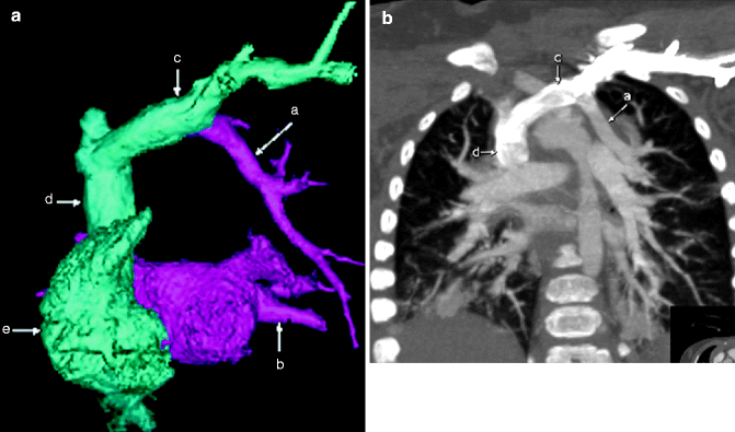
Fig. 5.6
Frontal projection 3D color-coded (a) and coronal MIP (b) images demonstrating a left upper lobe pulmonary vein (a) anomalously draining to the left brachiocephalic vein (c). The SVC (d) and right atrium (e) are mildly prominent. Notice that there is normal drainage of the left lower lobe pulmonary vein (b) to the left atrium
Supracardiac TAPVR Type 1
In the most common type of supracardiac TAPVR, the pulmonary veins form a common chamber connected to a vertical vein that in turn drains to the left brachiocephalic vein, which connects to the SVC. Occasionally, supracardiac TAPVR may connect to the right-sided SVC through a vessel that crosses the midline. Obstruction to the pulmonary venous blood flow may occur as the vertical vein crosses the bronchi or at its entry to the systemic vein. All types of TAPVR are left-to-right shunts that form obligatory right-to-left shunts through an ASD or patent ductus arteriosus after mixing with systemic venous blood. Thus, patients are cyanotic. This lesion may be isolated or associated with complex congenital heart disease and heterotaxy syndromes. All total anomalous pulmonary venous connections (TAPVCs) result in increased pulmonary blood flow.
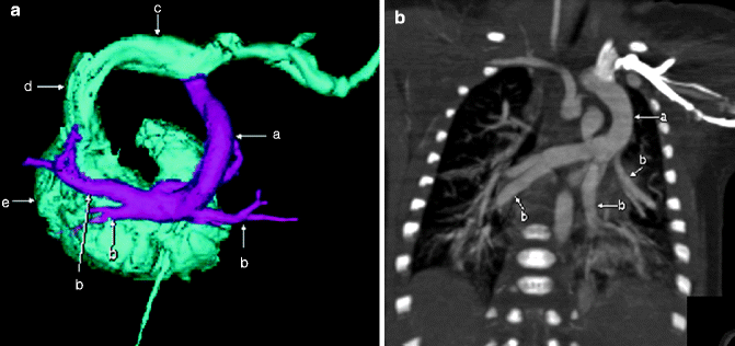
Fig. 5.7
Posterior projection 3D color model from CTA (a) and coronal MIP (b) images demonstrating multiple pulmonary veins (b) draining to a vertical vein (a), which connects to the brachiocephalic vein (c) and to the SVC (d) back to the systemic atrium (e)
Intracardiac TAPVR
Intracardiac TAPVR is also a left-to-right shunt that requires an obligatory right-to-left shunt through an ASD for adequate systemic cardiac output. Thus, patients may appear cyanotic. TAPVC draining into the coronary sinus is the second most common type of TAPVC; direct right atrial drainage is less common. Pulmonary venous obstruction is unusual.