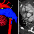
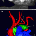
Fig. 9.1
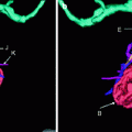
Posterior projection from a cardiac CTA image showing a typical left bronchus (A), which is elongated and is known as a hyparterial bronchus because the left main pulmonary artery (C) is draped over the proximal left bronchus (A). The right main bronchus (B) has a shorter course before giving off the upper lobe bronchus more proximally. Notice that the left main pulmonary artery (D) is not draped over the bronchus (B). This is known as an eparterial bronchus
Tracheal (Pig) Bronchus
A tracheal, or pig, bronchus is an anatomic variant (typically right) upper lobe bronchus originating from the trachea above the carina. Two types exist: displaced bronchus and supernumerary bronchus. This condition may cause persistent or recurrent right upper lobe pneumonias.
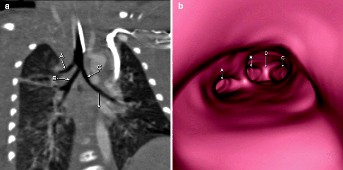

Fig. 9.2
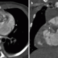
Coronal (a) and virtual bronchoscopy. Descending aorta (arrow) is shown. (b) Images demonstrating a right upper lobe accessory bronchus (A) arising from the trachea above the carina (D). The right bronchus (B) and left main bronchus (C) are shown
Airway Compression from Enlarged Pulmonary Arteries
Enlarged pulmonary arteries, when present, may be a frequent cause of airway compression in infants and small children with congenital heart disease. Enlarged pulmonary arteries frequently are seen in patients with congenital absence of the pulmonic valve, as observed in tetralogy of Fallot variants.
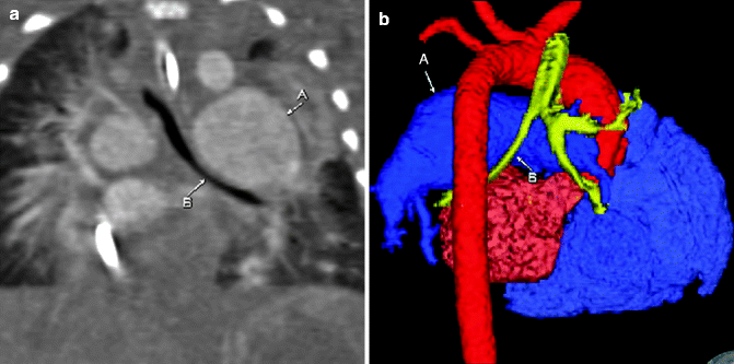

Fig. 9.3
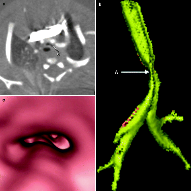
Enhanced coronal (a) and posterior projection color-coded three-dimensional (3D; b) images showing an enlarged left main pulmonary artery (A) with mass effect, resulting in moderate narrowing of the adjacent left main bronchus (B)
Tracheobronchomalacia
Tracheobronchomalacia is a tracheobronchial cartilaginous abnormality resulting in abnormally increased pliability and intermittent airway collapse. Dynamic examination, such as fluoroscopy or endoscopy, shows characteristic changes in airway caliber. Tracheobronchomalacia may be primary or secondary.
