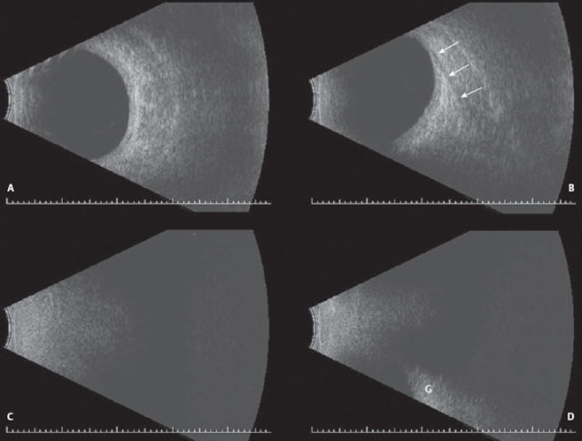
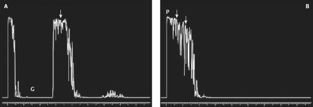
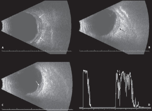
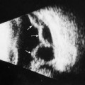
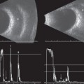
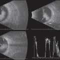
10 Over the last two decades in many practices, the role of ophthalmic ultrasound has advanced to include evaluation of the orbit. Aside from the retrobulbar optic nerves and extraocular muscles, which were discussed in previous chapters, evaluation of the orbital soft tissue, vasculature, and periorbital regions is now commonplace. Because ultrasound is a non-invasive procedure it can be used as the initial imaging tool prior to ordering more invasive tests or it can be used as an adjunct to other imaging studies when findings are not straight-forward. The same screening techniques that are used for evaluation of the globe can be employed for orbital screening; however, knowledge of the normal orbital anatomy is essential to obtain useful information. Transocular B-scan probe positions (vertical, horizontal, and oblique), longitudinal scans, and even axial scans can be used to evaluate the intraconal regions and the more posterior portions of the orbit. Paraocular B-scan probe positions (transverse, horizontal, and oblique) that by-pass the globe completely can be used to evaluate the more anterior regions of the orbits. All B-scan images provide the topographic features (location, size, shape, borders, and condition of the orbital bone) of the normal and abnormal orbital structures. Standardized A-scan provides the quantitative information (size, structure, reflectivity, consistency, vascularity, mobility, and sound attenuation) necessary to confirm the diagnosis. The normal orbital soft tissue is echogenic and highly reflective on both B-scan and standardized A-scan. Orbital disorders have highly variable echographic characteristics, so having a good understanding of any given patient’s medical history as well as knowing the histopathology of the various orbital lesions will improve the diagnostic ability of most echographers. The following images are a collection of some of the more commonly seen orbital lesions. We have included a few rare cases as well. Byrne SF, Green RL. Second Edition: Ultrasound of the Eye and Orbit. St. Louis: CV Mosby Yearbook; 2002 Dutton JJ, Byrne SF, Proia AD. Diagnostic Atlas of Orbital Diseases. Philadelphia: WB Saunders Company; 2000 Figure 10–2 Normal orbital A-scan (transocular/transverse). (A) Standardized A-scan with the sound beam directed through the globe (G) showing the highly reflective pattern from the normal orbital tissue (arrow). (B)Normal orbital A-scan (paraocular). Standardized A-scan with the sound beam bypassing the globe to show the highly reflective pattern from the normal orbital tissue (arrows). The probe (P) is placed directly over the tissue.
Evaluation of the Orbit
♦ Suggested Readings
![]()
Stay updated, free articles. Join our Telegram channel

Full access? Get Clinical Tree


