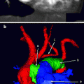
Fig. 6.1
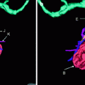
(a, b) Two axial views from a cardiac CTA demonstrating the typical anatomy of the right and left ventricles. (a) A prominent moderator band (B), which extends from the interventricular septum (A) to the free wall of the right ventricle. Notice how thin the membranous portion of the interventricular septum is compared to the more muscular portion (A). The adjacent left ventricular outflow tract is shown (D). The left ventricular wall typically is smooth and compacted (F) with papillary muscles (A). (b) The right ventricle demonstrates trabeculations (E) cordae tendinae(B). The mitral valve (C) regulates blood flow from the left atrium to the left ventricle, and the tricuspid valve (D) regulates blood from the right atrium to the right ventricle
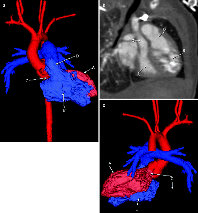
Fig. 6.2
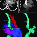
Frontal (a) and posterior (c) projection color-coded three-dimensional (3D) images, as well as an oblique view of the ventricles and outflow tracts (b), demonstrating a normal RVOT (D) originating from the more trabeculated right ventricle (B). The RVOT (D) is anterior to and to the left of the left ventricular outflow tract (LVOT; C). The LVOT arises from the more conically shaped left ventricle (A)
Membranous-Type VSD
Membranous-type VSD is also called perimembranous septal defect because the membranous septum is very small and the defect usually extends to the muscular septum. A membranous VSD is the most common type, accounting for 75–80 % of all VSDs. The defect lies in the outflow tract of the left ventricle immediately below the aortic valve. Tachypnea generally is the initial presenting symptom. Clinical symptoms depend on the size of the VSD. Small defects cause heart murmur with no other clinical symptoms. Moderate to large VSDs may cause tachycardia, diaphoresis, and failure to thrive. Congestive heart failure eventually may occur.
Echocardiography is the main diagnostic modality. In patients with large defects, chest radiography may show cardiomegaly with increased pulmonary vascularity. Kerley B lines from interstitial pulmonary edema may be seen in some cases. The location of the VSD is important for surgical repair.
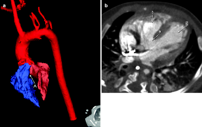

Fig. 6.3
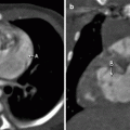
Color-coded 3D reconstruction (a) and contrast-enhanced axial cardiac CT image (b). A large perimembranous VSD (A) between the right (blue) and left (salmon) ventricles is demonstrated on the 3D reconstruction images. Note that the defect lies immediately beneath the aortic valve. The axial cardiac CT image (b) demonstrates a large area of communication (a) between the left (c) and right (b) ventricles, consistent with a perimembranous VSD
Intramuscular-Type VSD
Intramuscular-type, or trabecular-type, VSD accounts for 5–20 % of all VSDs. The defect is bordered by muscle within the trabecular septum, away from the cardiac valves. These VSDs vary in size. Surgical treatment depends on the size of the defect. Small defects may close spontaneously during the first 2 years of life, most within the first 6 months.
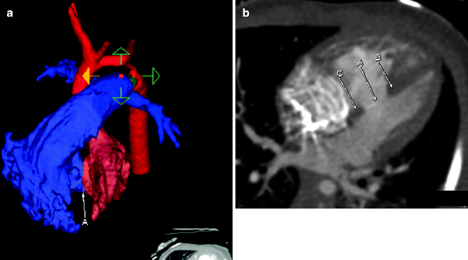

Fig. 6.4
Color-coded 3D reconstruction (a) and contrast-enhanced axial cardiac CT image (b). On the 3D reconstructed image, a VSD (A) is seen between the right (purple) and left (pink) ventricles. The corresponding axial CT image demonstrates the communication in the muscular interventricular septum (A). Notice that there is muscular tissue on the basilar (C) and apical (B) borders of the defect (A)
Multiple Intramuscular VSDs
Multiple intramuscular VSDs may have a “Swiss cheese” appearance when obliquely oriented through the interventricular septum. Surgical management is complex, with increased morbidity and mortality compared with other types of VSD.
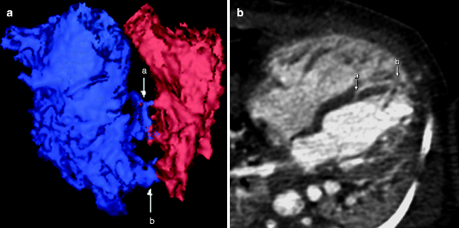

Fig. 6.5
Color-coded 3D reconstruction (a) and contrast-enhanced axial cardiac CT image (b). Two areas of communication are seen (a and b) between the ventricles involving the muscular septum
Posterior-Type VSD
Posterior-type, also known as AV septum– or inlet-type, VSD comprises 8–10 % of all VSDs. This defect, often associated with AV septal defect, occurs posterior to the septal leaflet of the tricuspid valve.
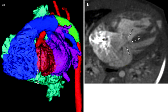

Fig. 6.6


Color-coded 3D reconstruction (a) and contrast-enhanced axial cardiac CT image (b). The 3D reconstructed image demonstrates abnormal communication between the ventricles posteriorly (C). The CT image demonstrates posterior-type VSD (C). Notice an associated septum primum defect (A) and a common AV valve (D). AV septal defects are associated with posterior-type VSD
Stay updated, free articles. Join our Telegram channel

Full access? Get Clinical Tree


