Fig. 3.1
The liver with its vasculobiliary skeleton; portal and hepatic veins (blue); hepatic arteries (red); bile ducts (green); gallbladder (GB); glissonian pedicle to segment 2 (GP2); glissonian pedicle to segment 3 (GP3); glissonian pedicle to segment 4 inferior (GP4i); glissonian pedicle to segment 4 superior (GP4s); glissonian pedicle to segment 5 (GP5); glissonian pedicle to segment 6 (GP6); glissonian pedicle to segment 7 (GP7); glissonian pedicle to segment 8 dorsal (GP8d); glissonian pedicle to segment 8 ventral (GP8v); glissonian pedicle to segments 5 and 8 (right anterior section) (GP5-8); glissonian pedicle to segments 6 and 7 (right posterior section) (GP6-7); inferior vena cava (IVC); left hepatic vein (LHV); middle hepatic vein (MHV); left glissonian pedicle (LGP); main glissonian pedicle (MGP); right glissonian pedicle (RGP); right hepatic vein (RHV); round ligament (RL); umbilical portion (UP)
The authors recommend liver explorations to start from the portal branches for the following reasons:
Get Clinical Tree app for offline access

1.
The portal bifurcation is generally the first structure encountered by positioning the probe on the liver’s surface since it is located relatively in the center, and can be visualized by positioning the probe in a relatively wide area of the liver surface which can be located along an ideal midline dividing the liver into an upper and lower portion, grossly passing through the edge between the segments 5 and 8 and 4 superior and inferior (Fig. 3.2a, b).
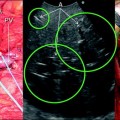
Fig. 3.2
Left (a) scan disclosing the main portal bifurcation and right (b) IOUS image; left portal vein (LPV); middle hepatic vein (MHV); right portal vein (RPV); portal branch to segments 5 and 8 (P5-8) (right anterior section); portal branch to segments 6 and 7 (P6-7) (right posterior section)
2.
Following the portal branches from their hilar origin to the periphery it is possible to explore all the segments without extensive movements but just titling the probe on the liver surface as recommended in Chap. 2 (Fig. 3.3a, b).
Fig. 3.3
Useful movements for liver exploration (as also shown in Chap. 2), involving up- and downward tilting (a) and axial rotation (b)
3.
Tilting the probe and following the portal branches from the central part to the periphery of the organ it is possible to identify and explore systematically all the sectional branches (Figs. 3.4a, b, 3.5a, b), and all the segmental and subsegmental portions of the liver (Figs. 3.6, 3.7, 3.8, 3.9, 3.10, 3.11, 3.12, 3.13, 3.14). One is then capable to locate any eventual lesion in relation to those branches and to recognize any eventual anomalous branching patterns (Figs. 3.15a, b, 3.16).
 Liver Transplantation from Living Donors
 Technical Tricks to Start Exploring the Liver with Ultrasound
 Diagnosis and Staging: Contrast-Enhanced Intraoperative Ultrasound (CEIOUS) Using Intravascular Contrast Agents
 Intraoperative Cholangio Ultrasound in the Study of the Biliary Tree
 Technological Requirements for Ultrasound-Guided Liver Surgery
 Liver Transplantation from Deceased Donors


Fig. 3.4
Left (a) scan, right (b) IOUS image; left portal vein (LPV); middle hepatic vein (MHV); right portal vein (RPV); portal branch to segments 5 and 8 (right anterior section) (P5-8); portal branch to segments 6 and 7 (right posterior section) (P6-7)
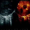
Fig. 3.5
Left (a) scan; right (b) IOUS image; right hepatic vein (RHV); portal branch to segments 5 and 8 (right anterior section) (P5-8); portal branch to segments 6 and 7 (right posterior section) (P6-7)
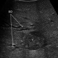
Fig. 3.6
a Scan, b intraoperative probe position, and c IOUS image; right portal vein (RPV); portal branch to segments 5 and 8 (right anterior section) (P5-8); portal branch to segment 5 (P5); portal branch to segment 8 (P8); tumor (T)
Fig. 3.7
Scan (a), intraoperative probe position (b), and IOUS image (c); inferior vena cava (IVC); right hepatic vein (RHV); portal branch to segment 8 (P8); portal branch to subsegment 8 dorsal (P8d); portal branches to segment 7 (P7)
Fig. 3.8
Scan (a), intraoperative probe position (b), and IOUS image (c); right hepatic vein (RHV); portal branch to segment 8 (P8); portal branch to segment 7 (P7)
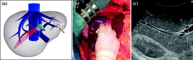
Fig. 3.9
Scan (a), intraoperative probe position (b), and IOUS image (c); kidney (K); portal branch to segments 6 and 7 (P6–7) (right posterior section); portal branch to segment 6 (P6); portal branch to segment 7 (P7)
Fig. 3.10
Intraoperative probe position (a), scan (b), and IOUS image (c); left hepatic vein (LHV); left portal vein (LPV); portal branch to segment 2 (P2); portal branch to segment 3 (P3); portal branch to segment 4 (P4); umbilical portion (UP)
Fig. 3.11
Scan (a), and IOUS image (b); portal branch to segment 4 inferior (P4i); umbilical portion (UP)
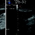
Fig. 3.12
Scan (a) and IOUS image (b); middle hepatic vein (MHV); portal branch to segment 4 superior (P4s); umbilical portion (UP)
Fig. 3.13
Scan (a) and IOUS image (b); portal branch to segment 3 (P3); umbilical portion (UP)
Related posts:
Liver Transplantation from Living Donors
Technical Tricks to Start Exploring the Liver with Ultrasound
Diagnosis and Staging: Contrast-Enhanced Intraoperative Ultrasound (CEIOUS) Using Intravascular Contrast Agents
Intraoperative Cholangio Ultrasound in the Study of the Biliary Tree
Technological Requirements for Ultrasound-Guided Liver Surgery
Liver Transplantation from Deceased Donors
Stay updated, free articles. Join our Telegram channel
Full access? Get Clinical Tree