EXTRACONAL ORBIT: ORBITAL PSEUDOTUMOR, NONINFECTIOUS INFLAMMATORY CONDITIONS, AND ACUTE AND CHRONIC INFECTIONS
KEY POINTS
- The imaging findings in inflammatory conditions involving the extraconal compartment may be nonspecific, but imaging patterns of involvement can aid in the differential diagnosis of causative pathology and direct medical decision making in many patients.
- Imaging can identify causative conditions for infections that can lead to improved outcomes if treated promptly.
- Imaging can identify complications such as detachments, retained foreign bodies, and compressive factors that may lead to improved outcomes if treated promptly.
Any orbital disease process might be approached by first establishing whether it is preseptal (Chapters 70 and 71) or postseptal (Chapters 57–60, 62, and 64). If postseptal, it should be established as a process arising primarily intraconally (Chapters 57–60), extraconally (Chapters 62 and 64), or transcompartmentally. If intraconal, it should be decided whether the condition is of optic nerve/sheath origin or arises from some other structure comprising or within the muscle cone. This chapter considers extraconal inflammatory processes and those arising extraconally and spreading across the orbital compartments and related spaces.
The noninfectious inflammatory condition of orbital pseudotumor and the myopathy associated with dysthyroid ophthalmopathy are by far the two most common inflammatory conditions that involve the extraconal fat. They most commonly involve both the muscle cone and extraconal space of the orbit. The effects of dysthyroid ophthalmopathy on the orbit are discussed in a dedicated chapter (Chapter 58) because they are so unique. Pseudotumor tumor is discussed in more detail in Chapter 57 since it is most commonly a disease of the muscle cone. The pathophysiology of those autoimmune processes and the relationship to other organ systems are discussed in Chapter 20. Other noninfectious inflammatory diseases may also be local or related to systemic disease.
The extraconal compartment is most often secondarily involved due to pathology involving the muscle cone or spreading from extraconal sources such as the sinonasal region (Chapter 87), osteomyelitis of the skull (Chapter 14), and the lacrimal gland (Chapter 65).
Infections, like noninfectious inflammatory conditions, may be local or related to systemic disease. Most of these conditions are due to secondary spread from conditions of the sinonasal region (Chapters 84–86) and lacrimal gland (Chapter 65). Spread of infections from intraconal (Chapters 55 and 57) and preseptal compartments (Chapter 70) is also common. Extraconal infections are also caused by penetrating trauma and surgery.
The spread patterns of these inflammations are discussed in the following Pathophysiology and Patterns of Disease sections.
ANATOMIC AND DEVELOPMENTAL CONSIDERATIONS
Applied Anatomy
The relevant anatomy of the bony orbit, muscle cone, and related neurovascular structures is discussed in detail in Chapter 44. In particular, the anatomy of the orbital periosteum, orbital septum, and attachments of the muscle cone at the orbital apex should be reviewed. The subperiosteal space within the extraconal, postseptal compartment lies between cortical bone and its adjacent periosteum. The postseptal extraconal space lies between the periosteum and muscle cone and contains fat and small nerves and vessels.
IMAGING APPROACH
Techniques and Relevant Aspects
The orbit, eye, and optic nerve and sheath are studied with computed tomography (CT) and magnetic resonance (MR) techniques and are described in detail in Chapters 44 and 45. Specific CT protocols by indications are detailed in Appendix A. Specific MR protocols by indications are outlined in Appendix B. Almost all studies to investigate extraconal inflammatory pathology are done with contrast.
Pros and Cons
Disease of and within the muscle cone is studied primarily with CT and magnetic resonance imaging (MRI). MRI is far more definitive in its rendering of the optic nerve and ocular effects of such disease and providing objective evidence of secondary optic nerve compromise. MRI is also far more sensitive than CT if there is any possibly related meningeal pathology. It detects intracranial involvement with these inflammatory processes and their intracranial complications much better than CT. MRI can also screen for related vascular complications and perivascular spread, especially in the cases of fungal disease.
CT typically is used in very acute situations, such as when an aggressive infectious process, perhaps with a sinonasal origin, is suspected or an emergent evaluation of possible associated tension orbit is required. It is also a first choice in patients too sick or for some other reason unable to complete a very high quality, free of motion artifact, MRI examination. CT is a reasonable secondary screening tool for identifying intracranial extension of disease and intracranial complications. Computed tomographic angiography is a good and perhaps preferred screening tool to search for vascular complications such as infectious aneurysms and cavernous sinus thrombosis.
SPECIFIC DISEASE/CONDITION
Orbital Pseudotumor/Idiopathic Orbital Inflammatory Syndrome and Noninfectious Inflammatory Disease
Etiology
Idiopathic orbital inflammatory syndrome (IOIS) is an acquired immune-based process that lacks systemic involvement.
Other noninfectious inflammatory diseases may also be local or related to systemic disease. The extraconal compartment is most often secondarily involved due to pathology involving the eye (Chapter 49) and structures within and comprising the muscle cone (Chapters 55 and 57). The granulomatoses and histiocytoses are infiltrating orbital processes that may present and appear identical to IOIS or lymphoma. These diseases are discussed in general in chapters on sarcoidosis (Chapter 18), Wegener granulomatosis (Chapter 17), and Langerhans histiocytosis (Chapter 19).
The features of autoimmune processes that may involve the extraconal space and the relationship to other organ systems are discussed in Chapter 20 in general and Chapter 58 for Graves dysthyroid orbitopathy.
Prevalence and Epidemiology
IOIS is a sporadic disease with no known predisposing factors.
The prevalence and etiology of the granulomatoses, histiocytoses, and autoimmune diseases that affect the muscle cone are discussed in the chapters on those entities as noted in the previous section.
Clinical Presentation
Pseudotumor almost always presents as a unilateral swollen and painful eye. There may also be visual loss and ophthalmoplegia (Figs. 57.3 and 57.5–57.7). It is occasionally bilateral. The other noninfectious inflammatory diseases are more likely to be bilateral than pseudotumor and the degree of pain and swelling less (Fig. 62.1).
Pathophysiology and Patterns of Disease
Although IOIS has several morphologic forms, its pathologic basis remains the same; only its distribution varies (Figs. 57.3–57.7). The disease most commonly involves the muscle cone and spreads both intraconally and extraconally; its more specific features are discussed in Chapter 57. Pseudotumor is unilateral in at least 85% of cases.1–3 In bilateral cases, it is often asymmetric. Bilaterality raises the likelihood of an alternative diagnosis of systemic disease such as lymphoma or sarcoidosis.
Brown superior oblique tendon syndrome is an adherence syndrome, which may be associated with IOIS or other inflammatory diseases or may be congenital. Imaging may show thickening of the tendon and trochlear region with edema in the surrounding fat.
The Langerhans cell histiocytoses may involve the orbit in children, usually as a bony lesion with secondary extraconal or transcompartmental orbital extension. The disease may permeate the orbital soft tissues. In the chronic or acute disseminated variants, spread within the optic sheath may be due to seeding from the subarachnoid space of the suprasellar cistern.
Granulomatoses most commonly involve the orbit as a result of spread from the sinonasal region; thus, extraconal involvement usually comes first. A transcompartmental pattern of spread is usually present at the periphery of the orbit where it borders the sinuses and/or nasal cavity. Sarcoidosis may present as a localized, infiltrating mass (Fig. 62.1).
Other very rare, with regard to their coming to imaging, infiltrating processes can involve the orbit and mimic IOIS and the granulomatoses and histiocytoses. Predominantly, dermatologic diseases such as Erdheim-Chester disease in adults and juvenile xanthogranulomatosis in children may secondarily involve the orbit. Necrobiotic xanthogranuloma, pseudorheumatoid nodules, amyloidosis, panniculitis, and other disease states must be distinguished on clinical grounds or by biopsy.
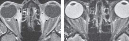
FIGURE 62.1. Magnetic resonance imaging study in a patient with orbital sarcoidosis. A, B: Contrast-enhanced T1-weighted fat-suppressed image (A) and T2-weighted fat-suppressed image (B) show both extraconal (arrows) and intraconal involvement with sarcoidosis (arrowheads).
Related posts:
 Preseptal Compartment: Acute and Chronic Infections and Noninfectious Inflammatory Conditions
 Intraconal Orbit: Tumors
 Hypopharynx: Developmental Abnormalities
 Tumors of the Submandibular Gland and Space and Tumorlike Conditions
 Masticator Space, Buccal Space, and Infratemporal Fossa Infections and Other Inflammatory Conditions
 Oral Cavity and Floor of the Mouth: Malignant Tumors
Stay updated, free articles. Join our Telegram channel

Full access? Get Clinical Tree


