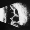
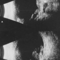
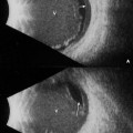
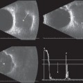
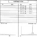
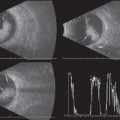
9 Clinicians have relied on their clinical assessment of patients and radiologic studies to diagnose and treat patients with disorders involving the extraocular muscles. Computed tomography (CT) and magnetic resonance imaging (MRI) easily show enlargement of the extraocular muscles if the enlargement is marked. Slight changes in the size of the muscles may go undetected with these tests, however, especially if the size of the section imaged is too large or an oblique cut is made, making the muscles look erroneously asymmetric or enlarged. Of course, CT involves exposure to radiation, and both CT and MRI are more expensive than ultrasound. Contact B-scan and standardized A-scan can be used to display the extraocular muscles from the inserting tendon at the globe to the muscle belly posteriorly. By B-scan the muscles display lower reflectivity than the surrounding orbital tissue because of the homogeneous nature of the muscle fibers. On standardized A-scan the extraocular muscles appear as a slight defect (area of lower reflectivity) within the highly reflective orbital fat. Although B-scan provides the topographic information (much like CT and MRI), the standardized A-scan provides precise measurements of the muscle thickness and information about the reflectivity that allows for differentiation of disorders that cause muscle enlargement. The normal values for the size of the extraocular muscles, as well as total volumes and acceptable differences between the muscle pairs have been established. Following the same prescribed B-scan examination techniques used for basic screening of the globe makes it relatively easy to look at the extraocular muscles in both cross-section (transverse) and radial (longitudinal) views. Once a single muscle has been imaged, it is recommended that the contralateral muscle then be displayed to determine any gross difference in thickness. The standardized A-scan can then be used to evaluate the muscle thickness and reflectivity. Echography can be useful in distinguishing between normal muscle patterns and disorders such as thyroid eye disease, myositis, and mass lesions involving the extraocular muscles. Examples of normal images and each of the various disorders are included on the following pages. Byrne SF, Gendron EK, Glaser JS, Feuer W, Atta H. Diameter of normal extraocular recti muscles with echography. Am J Ophthalmol 1991;112:706–713 Byrne SF, Green RL. Second Edition: Ultrasound of the Eye and Orbit. St. Louis: Mosby Yearbook; 2002
The Extraocular Muscles
♦ Suggested Readings
Related posts:
Stay updated, free articles. Join our Telegram channel

Full access? Get Clinical Tree


