Fair Game

Fair Game


Case 49


Comment


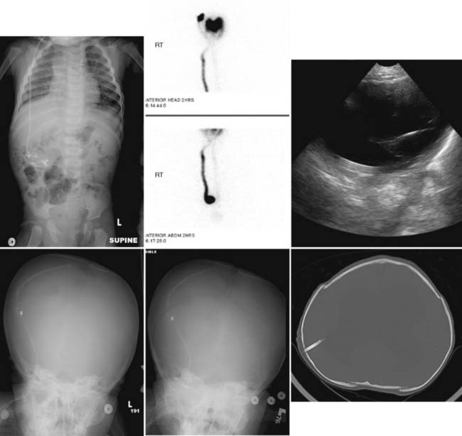
As better medical care ensures greater survival of premature infants with posthemorrhagic hydrocephalus, children with closed-head trauma, and children with brain tumors, the population of children with shunted central nervous system ventricles is growing. About 40% of shunts will eventually fail, either because of infection or mechanical dysfunction. Shunting in an individual patient generally begins with the VP approach. In a tiny, premature infant, a temporizing reservoir might be installed that drains to the subgaleal space; peritoneal drainage can be established when the infant gains sufficient size and cutaneous thickness. As the child grows, infection and fibrotic reactions can render the peritoneal space unusable. The neurosurgeon can attempt to establish drainage sites in the pleural spaces, the right atrium and superior vena cava, and even in the gallbladder.
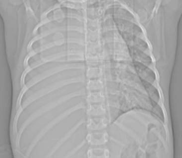

Complications of overshunting include subdural hemorrhage (occurring when the shrinking brain disrupts bridging vessels) and craniosynostosis, necessitating later surgical cranial expansion.


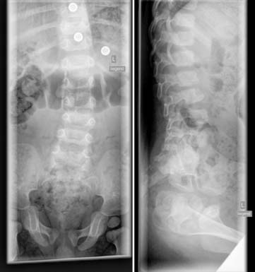
When the child with a shunt has a medical problem, it is axiomatic that the shunt is responsible until proven otherwise. Therefore the child generally receives a head CT scan to assess the ventricular size and position of the intracranial catheter. Next, plain films are taken that cover the pathway of the extracalvarial shunt tubing. It is essential to have comparison studies, both for the head CT scan and for the plain-film survey. Large ventricles on head CT scan may be the normal, balanced state for that patient, just as small ventricles may represent overshunting. Disconnection of the distal shunt tubing can be difficult to see when the valve is radiolucent: increasing space across the connection may be the only clue. Breakage most often occurs at the base of the neck—the point of maximum flexion stress. The tip should move around in the abdomen between studies. If it stays in one place, then it may be surrounded by fibrotic tissue and a high-pressure fluid collection might form. One exception to this is the recently perfected practice of inserting the shunt tip into the gallbladder; the tubing will remain closely coiled in the right upper quadrant, so clinical information is important to distinguish this from shunt fixation. The child can outgrow the shunt: as the torso lengthens, distal tubing is retracted from the peritoneum into the subcutaneous tunnel and can become obstructed. The chest and abdomen should also be assessed for signs of pneumonia, enteritis, or other medical problems that could cause the child’s symptoms.


Case 50


Comment


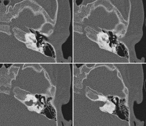
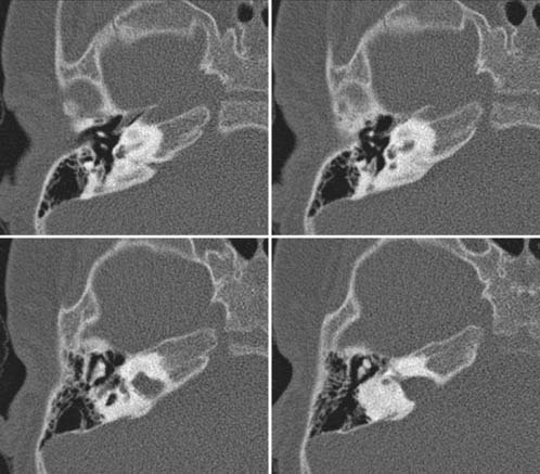
Membranous labyrinthitis with secondary ossification of the membranous labyrinth is the most common cause of acquired, bilateral sensorineural hearing loss (SNHL) in children. Ossification of the membranous labyrinth usually results from an infectious or traumatic insult. In children, typically SNHL occurs 2 to 18 months after an acute meningitis episode. The most frequent agents include S. pneumoniae and H. influenzae. The ossification occurs within the lumen of the otic capsule in response to the destructive, inflammatory process. The most common involved inner ear structure is the scala tympani of the basal turn. Clinically, a near complete deafness and vestibular malfunction may develop. Clinicians believe that extension of bacterial meningitis into the inner ear structures evolves along the cochlear aqueduct or internal auditory canal. Deafness after bacterial meningitis is observed in 5% to 20% of cases. Unfortunately, profound labyrinthine ossification is associated with a very poor prognosis.


Diagnosis is likely based on the combination of findings on high-resolution petrous bone CT and a matching positive, previous history of bacterial meningitis. CT may reveal an increased density of the cochlea and vestibulum. In early cases or in mild forms, the fibrosis and osteoid deposition may go undetected by CT. Magnetic resonance imaging (MRI) is especially helpful to rule out other reasons for acute deafness. In rare cases, during the acute phase, contrast-enhanced MRI may show an increased enhancement of the labyrinthine membrane. Prognosis has significantly improved because of the advent of cochlear implants.


Case 51


Comment


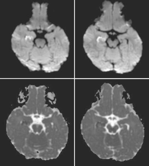
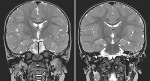
Prolonged or complex febrile seizures in early childhood may result in ischemic injury of the hippocampus with subsequent development of mesial temporal sclerosis. Mesial temporal sclerosis is associated with temporal lobe seizures. Febrile status epilepticus is defined as a seizure that lasts at least 30 minutes or a series of seizures lasting more than 30 minutes without interictal recovery in the setting of a febrile disease. Experimental animal studies showed that seizure-related injury is especially pronounced in the pyriform cortex and amygdale; however, the hippocampus is also involved. Acute MRI showed that the T2 hyperintensity of the hippocampal structures is preceded by a reduction of the apparent diffusion coefficient indicating cytotoxic edema. Histologically, the edema is rapidly followed by neuronal necrosis and tissue fragmentation. Provenzale and colleagues showed a strong correlation between the degree of hippocampal signal abnormality on acute imaging and subsequent hippocampal damage (respectively, development of mesial temporal sclerosis). Provenzale and colleagues also concluded that the identification of a marked signal abnormality after a prolonged febrile seizure might warrant the use of neuroprotective agents in the future.


Differential diagnosis may include a low-grade ganglioneuroma, astrocytoma, or a dysembryoplastic neuroepithelial tumor. High-resolution imaging of the temporal lobe usually allows the clinician to differentiate between acute postictal hippocampal swelling and a neoplasm or malformation.


Case 52


Comment


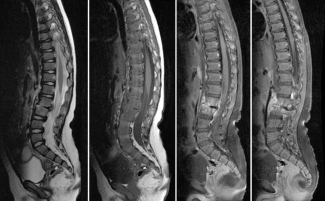
Childhood spondylodiscitis is a rare disease; the exact incidence is unclear. In spondylodiscitis, both the disk and the adjacent vertebral bodies are infected. In adults, rarely an isolated diskitis is seen. In children, because of the differences of the blood supply of the developing spine, an isolated diskitis may be observed more frequently. The clinical presentation is frequently nonspecific and may include refusal to walk or sit, back pain, inability to flex the lumbar spine, spinal scoliosis, and a loss of the normal lumbar lordosis. In addition, children may present with hip pain. Laboratory tests usually show nonspecific signs of infection, with a slight to moderately increased white blood cell count. Blood cultures are frequently negative. Initial radiographic studies frequently include an ultrasound of the hip as a first step, combined with radiography of the pelvis and spine. The radiography of the spine is frequently negative in the acute phase, because the typical radiographic findings usually do not appear until 2 to 3 weeks after onset of symptoms. Consequently, the diagnosis of a spondylodiscitis is usually delayed. Imaging findings on radiography include narrowing of the intervertebral disk, irregularity of the vertebral endplates, scoliosis, and possibly a widened paraspinal soft tissue line or an obscured psoas contour. Gas inclusions indicate abscess formation. MRI is the imaging modality of choice. The disk is usually narrowed and may show a strong contrast enhancement. In addition, T2-hyperintense bone marrow edema is seen in the adjacent vertebral bodies. The adjacent soft tissue may enhance, and an extension into the spinal canal with a possible epidural abscess should be excluded. In addition, a typical complication is an abscess formation in the adjacent paravertebral soft tissues, including a psoas abscess.


The cause of childhood spondylodiscitis remains unclear. Most frequently a generalized infection with sepsis is considered. Trauma, injections, or punctures may also be considered. S. aureus is the most frequently found organism in nontuberculous spondylodiscitis. Pneumococci, salmonella, and Escherichia coli infection are less common. Tuberculous spondylodiscitis is again on the rise and should always be considered. The therapeutic consequences are considerable. In nontuberculous spondylodiscitis, antibiotic agents and immobilization are usually recommended. If an epidural or paravertebral abscess develops, then abscess drainage is recommended. After the acute infection has resolved, frequently local fibrosis or bony ankylosis develops in the affected spinal segment.


Back pain may also result from spondylolysis; spondylolisthesis; traumatic injuries; disk degeneration and herniation; Scheuermann disease; tumors (primary, secondary, and hematogenous); and miscellaneous conditions (e.g., metabolic disorders, sickle cell disease, osteoporosis).


Refusal to walk, nocturnal waking with crying, and back pain in general in a child should always alert the clinician to the possibility of a spondylodiscitis.


Case 53


Comment


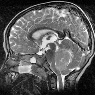
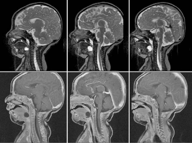
The Arnold-Chiari II malformation is named after Dr. Julius Arnold and Dr. Hans Chiari. This is a complex malformation that primarily involves the posterior fossa, which is too small. An extensive, complex combination of supra- and infratentorial malformations (and therefore findings) result from this condition. Arnold-Chiari II malformation is present in all patients with an open, nonskin-covered myelomeningocele. Clinicians believe that Arnold-Chiari II malformation results, at least in part, from leakage of cerebrospinal fluid from the open neural tube, which may prevent an adequate expansion of the rhombencephalic vesicle (believed to produce the small posterior fossa). Most patients are diagnosed prenatally. Functional outcome depends on the degree of hydrocephalus and the extent of the cerebral- and cerebellar-associated malformations. Magnetic resonance imaging is the best imaging modality to summarize all findings. Imaging findings include the small posterior fossa, a squeezed cerebellum and brainstem (resulting in a compression of the brainstem against the clivus), herniation of the cerebellar tonsils into the upper cervical spinal canal with kinking of the spinal cord, embracement of the brainstem by the cerebellar hemispheres, deformity (beaking) of the tectal plate, ascending herniation of the superior cerebellum through the tentorium cerebelli, supratentorial hydrocephalus with colpocephaly, thinning and/or dysgenesis of the corpus callosum, large massa intermedia, and a fenestrated hypoplastic falx cerebri with interdigitation of the mesial gyri. The occipital cortex may mimic polymicrogyria, which Barkovich has described as stenogyria.


Arnold-Chiari II malformation may be associated with syringohydromyelia in 50% to 70% of children. An increased incidence of diastematomyelia also exists.


Diagnosis is frequently made during the prenatal screening. Increased α-fetoprotein is highly suggestive of an open myelomeningocele. On prenatal ultrasound the downward displacement of the flattened and elongated cerebellum mimics a banana, whereas the inward depression of the frontal bones mimics a lemon.


Most children require a ventriculoperitoneal shunt to treat the hydrocephalus. Prognosis is variable and depends on the degree of hydrocephalus and associated malformations.


Case 54


Comment


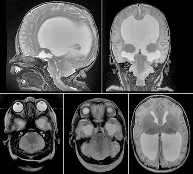
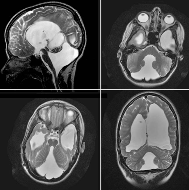
DWM is named after the neurosurgeon Walter E. Dandy and the neurologist Arthur E. Walker. A DWM is characterized by a cystic dilation of the fourth ventricle because of defective development of the anterior and posterior velum medullare. The vermis is hypoplastic or may be completely absent. A spectrum of DWM is known, and depending on the degree of malformation, different names are used. The more extensive anomalies are known as classic DWM or DW-variant; the less severe forms are known as Blake pouch cyst or mega cisterna magna. It should be kept in mind that not every cystic lesion within the posterior fossa is a DWM. Differential diagnosis includes a retrocerebellar cyst. In addition, DWM should be differentiated from a cerebellar hypoplasia or brainstem malformation, as well as from an infarcted cerebellum. The classic DWM is characterized by a cystic dilation of the fourth ventricle, usually with an absent choroid plexus, an upward rotated hypoplastic vermis, a large posterior fossa with elevation of the torcular herophili, and elevated straight sinus. The tentorium cerebelli may be hypoplastic. Frequently a supratentorial hydrocephalus occurs. Hydrocephalus does not have to be present at birth. In 60% of patients, associated findings are seen that include hydrocephalus, corpus callosum dysgenesis, migrational abnormalities (gray matter heterotopias, polymicrogyria, schizencephaly), occipital encephaloceles, and hydromyelia of the cervical cord. The neurocognitive development depends on the associated malformations; a majority of children will have a mental retardation. In addition, DWM is reported to be associated with cardiac anomalies and polydactyly. Magnetic resonance imaging (MRI) easily makes the diagnosis. A multiplanar MRI depicts all previously described findings. Differential diagnosis from a Blake pouch cyst and a mega cisterna magna may be challenging. A key feature is the identification of an intact vermis. In addition, DWM should be differentiated from acquired injuries of the cerebellum and vermis. Prognosis may differ significantly. Frequently, a DWM is diagnosed intrauterine by prenatal ultrasound examination. The fourth ventricle may have a typical keyhole appearance.


Case 55


Comment


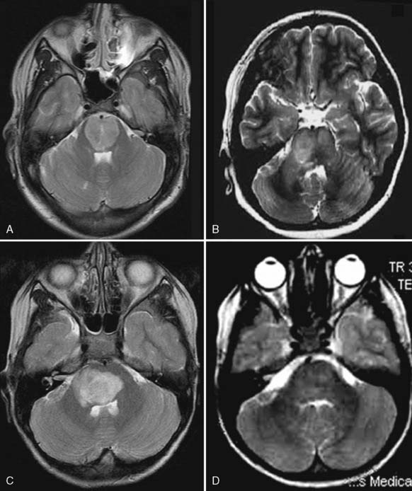
In children, many lesions may mimic primary brain tumors. A diffuse infiltrative brainstem glioma may have a long, benign course with only minimal clinical symptoms, significantly delaying diagnosis. In addition, symptoms may be attributed to various other causes. Diffuse infiltrative brainstem gliomas have a very slow growth and respect functional neurological center for a long time. Hydrocephalus occurs usually late during the course of the disease. Acute brainstem infarction is rare in children and presents with an acute onset of severe neurologic deficits, including various cranial nerve palsies and frequently dysregulation of respiration, hypoventilation, and dysregulation of temperature control. Prognosis is poor; most children die in the acute phase or may progress into a locked in syndrome. Acute brainstem infarction shows restricted diffusion on diffusion-weighted imaging and is usually symmetric in distribution. A thrombus within the basilary artery should be excluded. A mild mass effect may be observed because of the reactive edema. ADEM is an autoimmune reactive inflammation in response to a previous infection—frequently an upper respiratory tract infection. Lesions are frequently multifocal and involve the brainstem, basal ganglia, and thalami. Symptoms may be acute and diffuse. On imaging, lesions are T2 hyperintense, ill defined, and show a mild mass effect. On diffusion-weighted imaging, lesions usually show an increased diffusion compatible with vasogenic edema. If treated early, symptoms and lesions may resolve completely. UBOs are seen in neurofibromatosis and are not yet completely understood. These lesions are benign and usually without any clinical symptoms related to their location, extent, and size. Lesions are ill defined and T2 hyperintense, may show mild mass effect, and infrequently show a contrast enhancement. Lesions may change over time or disappear completely. Diagnosis is made in the setting of a neurofibromatosis. Multiple additional lesions may be seen throughout the brain.


It is essential to correlate imaging findings with the clinical findings to identify or rule out additional lesions and to use all current imaging modalities and functional sequences available to differentiate and characterize tumors from the various tumorlike lesions.


Case 56


Comment


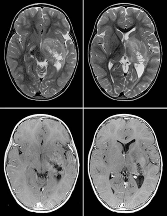
Cerebral astrocytomas are the most frequent supratentorial neoplasms in childhood. They represent more than 30% of all childhood supratentorial tumors. The majority of cerebral astrocytomas are fibrillary astrocytomas and may be of various degrees of malignancy; most of them are, however, low grade. Pilocytic astrocytomas are, with exception of a location within the hypothalamic-chiasmatic region, rare in a supratentorial location. The tumor may be solid, solid with necrosis, or solid with multiple cysts. Tumors are frequently in a deep location involving the thalamus or basal ganglia and may extend into the mesencephalon. On magnetic resonance image, low-grade variants are typically T2 hyperintense and show minimal or no contrast enhancement. Because the lesion diffusely infiltrates the adjacent brain, tumor borders are usually ill defined. The mass effect can be minimal or significant and complicated by obstructive hydrocephalus because of a compression of the adjacent ventricles. Higher-grade malignancies, especially a grade IV astrocytoma or glioblastoma multiforme, typically show a more extensive degree of contrast enhancement with big tumor cysts and extensive vasogenic edema. These lesions may be mistaken for abscesses. Diffusion-weighed imaging (DWI) is especially helpful in differentiating abscesses from tumor necrosis, because an abscess will be characterized by a restricted diffusion, whereas a necrotic tumor cyst will show an increased diffusion rate. Symptoms depend on the location of the tumor and may include seizures, focal neurologic deficits, or symptoms related to increased intracranial pressure, either by the tumor or by a complicating obstructive hydrocephalus. Currently, functional imaging, including DWI, diffusion tensor imaging, perfusion-weighted imaging, and 1H-MRS may help to characterize and determine the grade of malignancy of the tumor; however, a final histologic diagnosis is not yet possible. Tumor biopsy remains the gold standard for diagnosis. Therapy is determined by the location, age, and clinical symptoms. A deep, central location usually prevents surgical resection; a hemispheric location is more accessible for a surgical resection or at least tumor debulking followed by either chemotherapy and/or radiotherapy.


Case 57


Comment


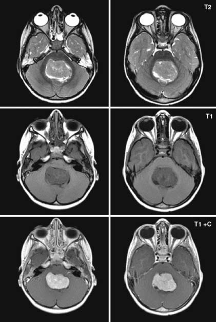
Medulloblastoma is, depending on the age and gender of the patient, one of the most frequent primary neoplasms of the posterior fossa. Medulloblastoma is especially frequent in boys in their first decade of life. Overall, medulloblastoma (25%) is second to pilocytic astrocytoma (35%). Brainstem gliomas (25%) and ependymomas (12%) are the third and fourth most frequent tumors of the posterior fossa in children. In total, these four variants represent 97% of all posterior fossa tumors. Medulloblastomas most frequently arise dorsal to the fourth ventricle either in the midline (vermis, 75% to 90%) or in a somewhat more lateral position (10% to 15%; also known as lateral medulloblastoma). Consequently, the fourth ventricle is pushed anteriorly and serves as an anterior tumor border. The compression of the fourth ventricle may result in an obstructive hydrocephalus. Clinical symptoms are either related to the obstructive hydrocephalus or to local tumor infiltration and may include ataxia, gait disturbance, nausea, vomiting, and headaches. Medulloblastomas have a high cellularity and are consequently dense on computed tomography. On magnetic resonance imaging the lesions can be differentiated from ependymomas, which are typically located within the fourth ventricle (in contrast to the medulloblastomas, which are primarily located dorsal to the fourth ventricle). Medulloblastomas usually display an ill-defined dorsal border because of infiltration of the adjacent vermis or cerebellar hemispheres; they are T1 hypointense to isointense and T2 isointense or hyperintense. Contrast enhancement may be strong but is occasionally absent. Cerebrospinal fluid (CSF) metastases may occur when the tumor has invaded the fourth ventricle. Tumor metastases may be seen within the third ventricle and lateral ventricles or along the spinal cord. A preoperative work-up should include the entire spinal axis. Prognosis depends on the residual tumor bulk after neurosurgical resection. The smaller the residual component, the better the prognosis. The residual tumor component is even of higher prognostic significance than the initial tumor size. In addition, the histology, immunohistochemistry, and neurogenetic results will also help to determine prognosis. Adjuvant treatment is decided by the combined information collected by imaging, immunohistochemistry, neurogenetic analysis, and residual tumor bulk after neurosurgery. Prognosis significantly improved in the last decade, with good long-term prognosis in most cases.


Case 58


Comment


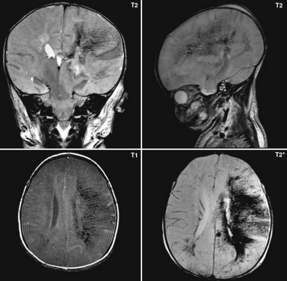
The germinal matrix is a highly perfused cell layer located along the ventricles from which the neurons originate that migrate toward the cerebral cortex. The germinal matrix is characterized by a high metabolic activity and is vulnerable for focal hemorrhages, especially in the preterm child. During ongoing development and maturation, the germinal matrix will progressively involute and finally disappear completely. Consequently, the risk for developing a GMH decreases with progressive gestational age.


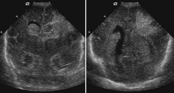
GMHs are classified into four grades. Grade I is defined as a focal hemorrhage confined to the germinal matrix. In grade II the hemorrhage will rupture into the ventricles that are not widened, whereas in grade III GMH the ventricles will be widened. This hydrocephalus is believed to result from focal adhesions along the ventricles, especially at the sylvian aqueduct and the outlets of the fourth ventricle, as well as by obliteration of the pachionic granulations. GMH grade IV has previously been defined as a GMH with extension into the adjacent hemispheric white matter (grade IV GMH is believed to represent a venous ischemia of the periventricular white matter because of compression and/or thrombosis of the deep, subependymal venous system by the hemorrhage). Secondary hemorrhagic conversion may complicate the hemorrhage. Clinically, grades I and II GMH may go undetected or may result in a focal seizure. Grade III is evident because of an increasing head circumference and neurologic instability. The development of a hydrocephalus is the major complication of GMH. Obstructive hydrocephalus may require frequent cerebrospinal fluid (CSF) punctures or even placement of a ventriculoperitoneal shunt. Diagnosis is usually made by transfontanellar ultrasound. GMH grade I is characterized by a focal, hyperechoic subependymal lesion along the ventricles (most frequently at the caudothalamic groove). Grade II hemorrhage is seen as a larger GMH in which blood products may cover the choroid plexus, resulting in a CSF-blood sedimentation level in the dependent parts of the ventricles or by a hyperechoic lining of the ventricles. Grade III is easily recognized by the enlarging ventricles. In grade IV hemorrhage, a hyperechoic signal is seen within the periventricular white matter (frequently in a fan-shaped pattern that follows the distribution of the venous drainage of the white matter into the deep venous system). A focal hemorrhage is seen as a focal hyperechoic mass lesion within the ischemic white matter. MRI is helpful for a better delineation and estimation of the degree of injury to the white matter. Functional sequences may give important information about functional outcome and prognosis. Follow-up examinations of GMH are usually done by serial bedside head ultrasound examinations. Color-coded duplex sonography with spectral analysis of the arterial flow profile with estimation of the resistive index may give important, indirect information about the intracranial pressure and the amount of brain edema. Serial, routine head ultrasound examinations are especially useful in children who are sedated and relaxed because of, for example, extracorporeal membrane oxygenation in heart failure.


Case 59


Comment


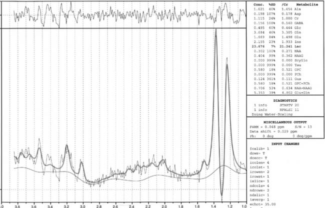
HIE is a very unfortunate combination of hypoxic injury to the brain because of hypoventilation and simultaneous ischemic injury to the brain (because of hypoperfusion resulting from, for example, cardiac arrest). HIE is a devastating injury that can occur intrauterine, perinatally during birth, or postnatally. HIE may also occur in near sudden infant death syndrome, after drowning, and in children with congenital heart diseases complicated by a sudden cardiac arrest. Depending on the age of the child (preterm versus term), the degree of hypoxia-ischemia, and the duration of hypoxia-ischemia, the distribution and severity of brain injury will vary. Preterm children with perinatal HIE will have injury to the periventricular white matter, which can evolve into periventricular leucomalacia (PVL), whereas term babies typically infarct their basal ganglia and thalami. Depending on the severity and duration of hypoxia-ischemia, various overlapping combinations of injury may be encountered. In addition, complicating germinal matrix hemorrhages may be seen in preterm neonates. Transfontanellar ultrasound is believed to be limited in the early detection of HIE. High-end ultrasound examination with combined color-coded duplex sonography and spectral analysis may, however, identify brain edema by measuring the resistive index. In addition, ultrasound may exclude other causes for the observed neurology. Neonates with severe HIE may be floppy, lethargic, have a low Apgar score, or may present with seizures. In older children with HIE, decreased consciousness, hypoventilation, hypothermia, or focal neurologic deficits may be observed. Magnetic resonance imaging (MRI), including DWI and quantitative proton magnetic resonance spectography (1H-MRS), give important functional data about the degree of injury. DWI can reveal ischemic injury to the brain before conventional MRI (T1– and T2-weighted imaging) shows pathologic condition. 1H-MRS may show increased concentrations of lactate within the brain, indicating anaerobic metabolism, while a decreased concentration of N-acetylaspartate (NAA) and creatine indicates neuronal cell injury and energy failure. On conventional MRI a reduced corticomedullary differentiation with swelling and increased T2 hyperintensity of the white matter are the most striking findings. Depending on the mechanism of injury, the basal ganglia and/or thalami may also be swollen. In neonates the T1-hyperintense and T2-hypointense signal of the myelinated white matter tracts in the posterior limb of the internal capsula (PLIC) have proven to be of prognostic value for outcome. If the PLIC signal is lacking, prognosis is poorer. In addition, intracortical T1-hyperintense foci within the central region indicate HIE with intracortical petechial hemorrhages. The intramedullary veins may also be prominent because of increased venous pressure or thrombosis. Functional MRI is especially helpful in examining children with HIE because it gives valuable, early information before conventional MRI shows the pathologic condition. Consequently, neuroprotective treatments can be started earlier and more selectively.


Case 60


Case 61


Comment


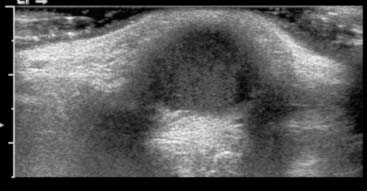
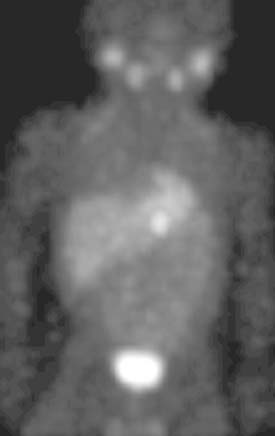
The thyroid begins to form in the fourth week of fetal life as a pit in the base of the tongue, at the junction of the proximal one third (derived from the third branchial arch) and the distal two thirds (derived from the first branchial arch). As the fetus enlarges, the pit deepens, forming a duct in the midline—the thyroglossal duct—with the developing thyroid at its distal tip. Ultimately this duct passes inferiorly through the base of the tongue; then it passes anteriorly to the surface of the neck, passing superior and anterior to the hyoid bone and anterior to the thyroid and cricoid cartilages. The rapidly enlarging bilobed thyroid reaches its place in the lower neck by the end of the seventh week; usually by that time the duct above it has disappeared, persisting only as the vestigial pit in the tongue—the foramen cecum—and occasionally as the pyramidal lobe of the thyroid, projecting upward from the isthmus. However, remnants of the duct can persist as cysts anywhere along its pathway. Similarly, remnants of thyroid can be left behind as solid masses.


The complexity of the duct’s path complicates its imaging workup. Cysts in the anterior neck are easily studied using ultrasound. However, cysts or masses in the base of the tongue may not be accessible, and computed tomography or MRI might be necessary. In some cases the thyroid is completely arrested in its descent, and the mass may actually represent the patient’s entire complement of thyroid tissue. Therefore ultrasound of the thyroid to establish its normal configuration is mandatory before surgery is contemplated. If thyroid tissue is not in its normal place, then nuclear medicine thyroid scan must be done to determine its presence and location.


Because the path of the thyroglossal duct crosses the territories of the second, third, and fourth branchial arches, the differential diagnosis of a mass in the base of the tongue or anterior neck must also include cystic remnants of these structures. Generally these structures are off-midline and more lateral.


Case 62


Comment


Rhabdomyosarcoma is the most common soft tissue sarcoma in childhood. The name comes from the Greek words rhabdo, meaning rod shaped, and myo, meaning muscle. The tumor arises from primitive muscle cells; tumor cells are usually positive for desmin, vimentin, myoglobin, actin, transcription factor myoD, and elements of differentiated muscle cells. The incidence is four to seven per 1 million children younger than 15 years old in the United States (or about 250 cases annually). Two thirds of the patients are younger than 10 years old. The boy-to-girl ratio is 1.2:1.4 to 1. Most cases occur sporadically and the cause is unknown. However, a pattern of familiar cancer exists, including osteosarcoma and rhabdomyosarcoma in the Li-Fraumeni syndrome, which is seen in children with first-degree relatives with adrenocortical carcinoma, breast cancer, or other tumors that occur before the age of 45 years. Tumor-suppressor gene p53 mutations are associated with this syndrome.


Rhabdomyosarcoma can occur anywhere in the body but does not arise primarily in bone. The most common sites are the head and neck (28%), extremities (24%), and genitourinary (GU) tract (18%). Other sites include the trunk (11%), orbit (7%), retroperitoneum (6%), and other sites in less than 3% of patients. The botryoid variant of embryonal rhabdomyosarcoma arises in mucosal cavities, such as the bladder, vagina, nasopharynx, and middle ear. Lesions in the extremities are most likely to have an alveolar type of histology. Metastases are found predominantly in the lungs, bone marrow, bones, lymph nodes, breasts, and brain.


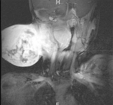
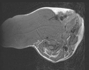
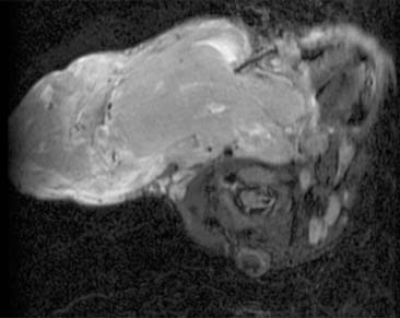
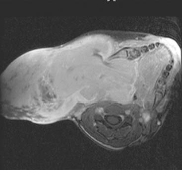
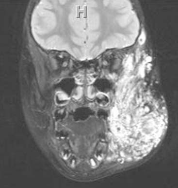
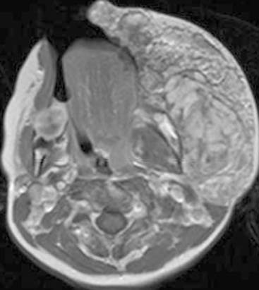
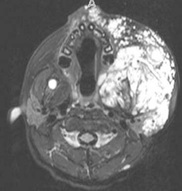
The images shown in this case are of an unfortunate boy who was noted to have an egg-sized mass 3 months before this MRI was obtained. Initial treatment with antibiotic agents had had no effect on the mass, and biopsy 1 month later showed embryonal rhabdomyosarcoma. The mass grew rapidly. The family sought several medical opinions, and he was first treated with herbal supplements. He never received conventional therapy and died at home 8 months after the MRI examination.


Case 63


Comment


Subglottic stenosis is one of the most common causes of airway obstruction in infants and children. It is the second most common cause of stridor in infants and the most common laryngotracheal anomaly requiring tracheostomy in children younger than 1 year old. It is the most common serious long-term complication of endotracheal (ET) intubation in neonates. Subglottic stenosis may be categorized as congenital or acquired. The diameter of the normal subglottic lumen is 4.5 to 5.5 mm in a full-term neonate and approximately 3.5 mm in a preterm baby. A subglottic airway diameter of 4 mm or less in a full-term infant or 3 mm or less in a premature infant is considered narrow and consistent with a diagnosis of subglottic stenosis. Subglottic stenosis is considered congenital when no other apparent cause of the stenosis exists. Congenital subglottic stenosis implies that a child is born with a small laryngeal lumen and that trauma of intubation or another cause did not contribute to the stenosis. After intubation, it is difficult to distinguish congenital from acquired stenosis. A congenitally malformed larynx leads to respiratory distress that may require intubation. Inflammation and scarring may occur even though an age-appropriate size of ET tube was used. Therefore the true incidence of congenital subglottic stenosis is difficult to determine. The majority of cases of subglottic stenosis are acquired and most commonly associated with ET tube intubation and many other factors, including laryngopharyngeal reflux, infection, and the associated inflammatory response. ET tubes may cause pressure injury to the glottis, whereas tracheotomy tubes may cause severe stomal stenosis in the trachea or infraglottic region.


Plain films may be the first modality of choice; however, thin-slice CT images provide better anatomic detail and allow for 3D reconstructions of the larynx and airways. Short scan time allows for scanning of the child without intubation or sedation, making it more preferable than magnetic resonance imaging.


Acquired subglottic stenosis may present years after ET intubation. Surgical correction of subglottic stenosis aims to provide an adequately enlarged lumen while preserving vocal quality and airway protection. Treatment success is predicted on a thorough preoperative evaluation and tailoring the repair to address the severity and location of the individual lesion.


Differential diagnosis is broad and includes—but is not limited to—tracheomalacia, laryngomalacia, laryngeal cleft, vascular compression, teratoma, vascular anomalies (e.g., hemangiomas, lymphatic malformation), and recurrent respiratory papillomatosis.


Case 64


Comment


Venous malformations represent a subcategory of vascular malformations that are congenital malformations of vessels that are not true neoplasms.


In 1982, Mulliken and Glowacki published a landmark article proposing characterization of vascular anomalies based on biologic and pathologic differences. This classification system clarified the misconceptions and misnomers that had been developing within the group of vascular anomalies during the past 200 years. Vascular anomalies are divided in two major groups: (1) hemangiomas (true vascular tumors) and (2) vascular malformations (including venous malformations, lymphatic malformations, capillary malformations, mixed type of venolymphatic malformations, arteriovenous malformations, and arteriovenous fistulas).


Venous malformations represent dysplasia of small and large venous channels. They generally present as painful soft tissue masses, sometimes with a bluish hue. They may bleed and result in cosmetic problems. Venous malformations are commonly diagnosed at birth but may become apparent at any age. They may suddenly enlarge secondary to hemorrhage or hormonal influences.


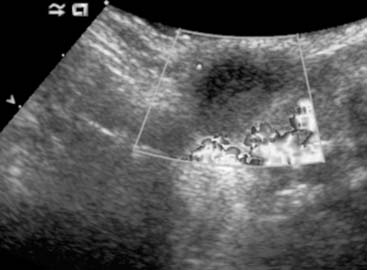
Radiographs may show a soft tissue mass. Computed tomography is typically not used in the diagnostic workup. Ultrasound can show mixed echogenicities within tangles of hypoechoic tubular structures. Color Doppler sonography does not show any arterial flow. Venous malformations reveal increased T2 signal and appear as channel-like serpiginous areas. They show avid contrast enhancement and can have calcifications that appear as signal voids (phlebolith). Venous malformations are infiltrative lesions crossing multiple soft tissue planes involving subcutaneous fat, bone, neurovascular bundles, or even viscera. Magnetic resonance imaging is preferred for monitoring the disease, whereas ultrasound is commonly used to rule out deep venous thrombosis after treatment.


Differential diagnosis includes hemangiomas and lymphatic malformations. Hemangiomas are soft tissue mass lesions that show arterial flow within their parenchyma; venous malformations do not have any arterial flow. Venous malformations typically present with tubular serpiginous T2-bright, avidly enhancing mass lesions, whereas lymphatic malformations are generally cystic lesions that are again T2 bright but do not enhance except for the wall of the cysts or septations.


Aspirin may be used to avoid thrombosis. Elastic compression garments are recommended. The primary modality of treatment is percutaneous sclerosis, such as direct injection with ethanol or other sclerotic agents under fluoroscopy or ultrasound guidance (performed under general anesthesia). Potential complications of percutaneous treatment of vascular malformations include skin necrosis, nerve damage, extremity swelling, muscle atrophy, and deep vein thrombosis. Often this is a life-long problem, with treatment aimed at reducing symptoms rather than eliminating disease. A multidisciplinary approach facilitates appropriate diagnosis, management, and treatment of these cases.


Case 65


Dec 21, 2015 | Posted by in PEDIATRIC IMAGING | Comments Off on Fair Game

Full access? Get Clinical Tree

Get Clinical Tree app for offline access