Chapter 12. Female Pelvis
Patient Preparation
• Full bladder: 32 ounces of clear fluid before the examination; no voiding until after the examination is completed. Alternative: no preparation, TA imaging followed by endovaginal EV imaging.
Equipment and Technical Factors
• Curved linear, vector, linear array (for abdominal wall imaging), and EV transducers are used. The EV transducer should be adequately sheathed for the examination and appropriately disinfected after each use. Published guidelines for disinfection of endocavitary transducers are available. In addition, manufacturer specifications should be followed to avoid damaging the transducer.
• Color Doppler imaging can be used to distinguish vascular from nonvascular structures.
Imaging Protocol
• Longitudinal axis images through the medial, mid, and lateral aspects of the uterus and both adnexa. The endometrial thickness should be evaluated with the endometrium perpendicular to the beam (may be done by TA imaging, but EV imaging is more accurate).
• Transverse axis images through the cervix, body, and fundus of the uterus and both adnexa.
• Longitudinal and transverse axis images of the ovaries.
• EV imaging may be performed with the patient in the lithotomy position (gynecologic examination table with stirrups is preferred; a covered support wedge may be used to elevate hips) or the Sims position (useful when the patient is obese or cannot lie supine).
• Doppler evaluation of the ovaries will demonstrate variation in blood flow with menstrual cycle (resting versus ovulating ovary).
• Bimanual technique (external pressure over pelvic area) may place uterus and/or ovaries into the scan plane.
Measurements
Uterus (nulliparous)
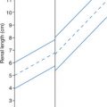
• Length: 7.0 cm
• Width: 5.0 cm
• Thickness (AP): 3.0 cm
Uterus (multiparous)
• Approximately 2.0 cm larger in all three dimensions
Uterus (postpartum)
• Enlarged uterus should involute to multiparous size within 4 to 8 weeks after delivery
Uterus (postmenopausal)
• Length: 3.0-5.0 cm
• Width: 2.0-3.0 cm
• Depth (AP): 2.0-3.0 cm
Endometrial thickness
• Menstruating: 4.0-14.0 cm
• Postmenopausal: <5.0 mm
Ovary
• Length: 2.5-5.0 cm
• Postmenopausal: 5.8 cm3
• Thickness (AP): 0.6-2.2 cm
• Width: 1.5-3.0 cm
Ovarian volume 0.523 (L × W × D)
| Female Pelvis | |||
|---|---|---|---|
| Sonographic Finding(s) | Clinical Presentation | Differential Diagnosis | Next Step |
| Anechoic structure(s) in cervix | Asymptomatic | Nabothian cyst(s) | Document size and location |
Enlarged, bulky uterus with distinct mass(es) noted Possibly heterogenous echotexture without identification of specific mass(es) Possible irregular border Hypoechoic, hyperechoic, heterogenous, or complex mass(es); small to very large in size Possible calcification within mass(es) Possible displacement of endometrial canal Hydronephrosis/hydroureter may be noted | Uterus enlarged on manual examination Asymptomatic to mild symptoms Common complaints: feeling of pelvic “fullness” or pressure, back pain, urinary incontinence, painful periods | Leiomyoma (fibroid/myoma) Intramural Subserosal Pedunculated All types may demonstrate necrosis and degeneration All types may enlarge during pregnancy and regress after menopause Adenomyosis | Intramural fibroids may displace endometrial canal without change in overall uterine shape Subserosal fibroids distort contour of uterus even when small Pedunculated fibroids may not demonstrate connecting stalk Evaluate urinary bladder for impingement by uterus |
| Normal size uterus with focal thickening or distortion of endometrium | Heavy bleeding during periods, may interfere with fertility Labs: decreased hematocrit; anemia (if bleeding is heavy and prolonged) | Leiomyoma Submucosal Endometrial polyp Intramural fibroid | Sonohysterogram to reveal true endometrial thickness and confirm presence of submucosal fibroid or polyp 3D imaging may be helpful Fibroid may prolapse into vagina |
| Ovarian cyst, <2.5 cm in diameter | Asymptomatic | Dominant follicle (menstruating women) Postmenopausal woman: suspicious for ovarian carcinoma | Serial sonograms should demonstrate change in cyst with menstruation |
Ovarian cyst, 3.0 cm to 20.0 cm Possible internal echoes | Asymptomatic Pelvic pain if cyst is large Hemorrhage may cause fever, pain Dysfunctional uterine bleeding if cyst produces hormones | Follicular cyst | Internal echoes may indicate hemorrhage Large cyst may cause ovarian torsion |
| Uterine body and fundus are the same size as cervix | Asymptomatic | Menopausal woman: normal atrophy of uterus Prepubertal uterus | Size and appearance of ovaries should correlate with uterus |
Enlarged uterus with bulbous fundus Uterus is hypoechoic and heterogenous Anterior or posterior wall may be eccentrically enlarged Small cysts in myometrium Multiple areas of attenuation | Painful vaginal bleeding Heavy bleeding | Adenomyosis Diffuse/focal Leiomyoma Myometrial contraction Endometrial carcinoma | Most commonly found in multiparous women |
Rapid enlargement of fibroid Hydroureter hydronephrosis may be noted | Possible rapid increase in abdominal girth | Leiomyosarcoma | Evaluate urinary bladder for impingement by uterus |
| Bulky, enlarged cervix | Asymptomatic Labs: positive PAP smear | Cervical carcinoma Cervical myoma | Evaluate urinary bladder for impingement by cervix |
| Two uterine horns, two cervices, two vaginas | Asymptomatic Uterus enlarged on manual examination Heavy periods | Uterus didelphys | Associated with renal anomalies and pregnancy complications |
| Two uterine horns noted (two endometria) | Asymptomatic Uterus enlarged on manual examination Heavy periods | Uterus bicornis | Associated with renal anomalies and pregnancy complications |
Two endometrial stripes are noted
Related posts:Stay updated, free articles. Join our Telegram channel
Full access? Get Clinical Tree


| |||