Chapter 13. First Trimester Pregnancy
Patient Preparation
• Preparation is similar to that for the female pelvis; however, an extremely full bladder may compress the gestational sac. Alternative: no preparation, TA imaging followed by EV imaging may be done.
Equipment and Technical Factors
• MI and TI settings must comply with the ALARA principle; pulsed-wave Doppler imaging should not be used during the first trimester to obtain an EHR because of the high intensity than can be focused on the developing heart.
• If there is difficulty locating the embryonic heart with 2D imaging, color or power Doppler imaging may be used sparingly to locate the heart for M-mode cursor placement.
• Doppler imaging may be used in the evaluation of adnexal pathologic conditions.
Imaging Protocol
• Longitudinal axis images through the medial, mid, and lateral aspects of the uterus should include the gestational sac; include images of both adnexa and ovaries.
• The gestational sac should be evaluated for the presence of absence of an embryo, early placenta, and membranes (EV imaging provides the best resolution).
• Transverse axis images through the cervix, body, and fundus of the uterus should include the gestational sac; include images of both adnexa and ovaries.
• Appropriate measurements are performed: MSD and CRL of the embryo; the yolk sac diameter may be obtained.
• Compare measurements obtained during the sonographic examination with the date of the patient’s last menstrual period and the estimated delivery date.
• Near the end of the first trimester, fetal biometry may be obtained.
Measurements
Gestational Sac
• Calculate the MSD (L + W + D)/3; correlate with patient’s dates; accuracy is +1 week.
CRL
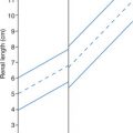
• Accuracy +3 days; refer to published charts (see Appendix A).
Yolk sac
• Maximum diameter: 5.0-6.0 cm
Nuchal translucency at 9–13 weeks (45.0 mm–84.0 mm CRL)
• <3.0 mm
• MSD and CRL measurements can be referenced to standard charts and recorded on a technical worksheet if software package is not available.
• The location and size of a uterine fibroid should be documented during each sonogram of the pregnancy.
| First Trimester | |||
|---|---|---|---|
| Sonographic Finding(s) | Clinical Presentation | Differential Diagnosis | Next Step |
No yolk sac noted in a gestational sac >10.0 mm (EV scanning) No embryo noted in a gestational sac >18.0 mm (EV scanning) Irregular sac with lack of decidual reaction Sac is low in uterus | Pregnancy “feels different”; diminished morning sickness and breast tenderness Vaginal bleeding Cramping Advanced maternal age Diabetes History of recurrent abortions Labs: positive β-hCG | Anembryonic pregnancy (blighted ovum) Normal intrauterine pregnancy Pseudosac (ectopic pregnancy) | Serial β-hCG and sonograms may be done May require surgical intervention to resolve |
| Yolk sac measures >6.0 cm with/without presence of embryo | Asymptomatic | Enlarged yolk sac | Associated with pregnancy loss |
Ovarian cyst, <4.0 cm with thick walls and internal echoes Increased flow noted with color Doppler imaging | Asymptomatic Hemorrhage may cause pain Labs: increased progesterone | Corpus luteum cyst | Should not persist after 16 weeks’ gestation May need serial scans to follow regression |
| Cystic area in posterior fetal brain noted at 8–11 weeks’ gestation | Asymptomatic | Normal rhombencephalon (hindbrain) | Precursor to fourth ventricle |
| Protrusion or bulge at umbilical cord insertion into embryo/fetus noted at 8–12 weeks’ gestation | Asymptomatic | Normal physiological gut/umbilical herniation
Related posts:Stay updated, free articles. Join our Telegram channel
Full access? Get Clinical Tree


| |