1 Flank Pain Flank pain is a common complaint and is often the primary symptom of ureteral stone disease. However, the differential diagnosis of flank pain is extensive and includes not only many renal etiologies but also nonrenal etiologies.
Retroperitoneal | Renal | Calculus disease |
|
| Pyelonephritis/abscess |
|
| Xanthogranulomatous pyelonephritis |
|
| Neoplasm/cyst |
|
| Vascular compromise |
| Nonrenal | Abdominal aortic aneurysm rupture |
|
| Retroperitoneal fibrosis |
|
| Retroperitoneal abscess |
|
| Retroperitoneal adenopathy |
|
| Retroperitoneal hemorrhage |
|
| Retroperitoneal neoplasm |
|
| Adrenal neoplasm with hemorrhage |
Intraabdominal | Diverticulitis |
|
| Perforated colon carcinoma |
|
| Pancreatitis |
|
| Cholecystitis |
|
| Appendicitis |
|
| Duodenal ulcer |
|
| Splenic infarction/abscess |
|
Musculoskeletal | Fibrositis |
|
| Iliac/spine osteomyelitis |
|
| Disk disease |
|
| Spinal metastases |
|
Miscellaneous | Münchhausen syndrome |
|
| Narcotic abuse |
|
The approach to the patient with flank pain begins with a thorough history and physical examination followed by appropriate laboratory studies. This information will help the clinician in consultation with the radiologist to determine which radiological procedures are indicated. Table 1–1 lists the renal and nonrenal causes of acute flank pain.1,2 Many of the disease processes listed in this table occur commonly, including renal calculus disease, acute pyelonephritis, and fibrositis (Table 1–2).1,2 Although many of the intraabdominal disease processes listed are also common, flank pain is an unusual manifestation. This chapter presents a brief summary of the important diagnostic features of these disease processes.
Differential Diagnosis
An abrupt increase in intraluminal pressure in the collecting system is the primary cause of pain from calculus disease. The severity of the pain is directly related to the rapidity with which the obstructing stone raises intraluminal pressure.3 The pain frequently radiates from the flank laterally into the abdomen and may be intermittent (renal colic) or steady. When the ureter is initially obstructed, there is an increase in the frequency of ureteral contractions causing colicky pain. Within a short time, ureteral contractions cease, and while the intraluminal pressure remains elevated, the pain takes on a more steady character.3 Nevertheless, the patient typically paces or writhes in bed, unable to find a comfortable position. The location of pain originating from the urinary tract has been carefully mapped out by McClellan and Goodell.3 The authors observe that if various parts of the renal pelvis and ureter are distended with small balloon catheters, the distention of the renal pelvis produces costovertebral angle pain. When the upper, middle, or lower ureter is distended, pain occurs in the flank, middle inguinal canal, or suprapubic region, respectively. Pain may also be referred from the upper or lower obstructed ureter to the ipsilateral scrotum in men or labia majora in women.3 Symptoms of bladder irritation may occur. Because of the autonomic innervation of the kidney and stomach by the celiac ganglion, it is not unusual for patients to experience nausea, vomiting, and abdominal distention due to ileus. Diaphoresis and tachycardia may also occur in response to the pain; however, fever is not usually present unless there is a superimposed infection and pyonephrosis. Urinalysis usually reveals microscopic hematuria; however, gross hematuria occurs in 10% of patients. If ureteral obstruction is complete, then hematuria may be absent.4
Common (100) | Uncommon (5–100) | Rare (0–5) |
Renal colic | Renal corticomedullary abscess | Renal cortical abscess |
Acute pyelonephritis |
| Perinephric abscess |
Fibrositis | Carcinoma of the kidney | Renal infarction |
| Diverticulitis | Renal vein thrombosis |
| Carcinoma of the colon with perforation | Adrenal neoplasm with hemorrhage |
| Duodenal ulcer | Iliac osteomyelitis |
| Acute cholecystitis | Retrocecal appendicitis |
| Abdominal aortic aneurysm rupture | Splenic infarction |
|
| Splenic abscess |
ªIncidence per 100,000 (approximate).
Acute pyelonephritis is characterized by the abrupt onset of chills and fever, usually > 100°F, in association with constant, dull flank pain and/or symptoms of cystitis, which include dysuria, frequency, and urgency. In some cases, only irritative bladder symptoms are present, and differentiating pyelonephritis from cystitis is difficult.5 The flank pain is usually less severe than that caused by stone disease and may be associated with muscle spasm and tenderness that intensify with movement. Nausea, vomiting, diarrhea, and abdominal pain may also occur. Laboratory findings show a leukocytosis with a neutrophil predominance. Urinalysis reveals numerous white blood cells and bacteria. Leukocyte casts and a specific type of cast characterized by the predominance of bacteria within the cast matrix are also frequently present in the urine.5 Urine cultures usually grow Escherichia coli. When pyelonephritis occurs in the setting of stasis, calculi, pregnancy, diabetes mellitus, or a neurogenic bladder, patients are pre-disposed to abscess formation.5 If renal abscess develops from a hematogenous source, then urine cultures will frequently be negative.
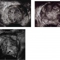
An uncommon infectious process of the kidney, xanthogranulomatous pyelonephritis, predominantly affects women between the fifth and seventh decades of life. Patients frequently present with nonspecific constitutional symptoms such as malaise, low-grade fever, and flank pain, which have usually been present for months to years.6 On physical examination, there is costovertebral angle tenderness and frequently a palpable flank or abdominal mass. Laboratory findings reveal an elevated erythrocyte sedimentation rate, anemia, and leukocytosis. Urinalysis shows pyuria, proteinuria, and occasionally hematuria. Escherichia coli and Proteus mirabilis are often cultured from the urine, although urine cultures may be negative in up to one third of patients if there is complete obstruction of the kidney.6 An interesting syndrome of reversible hepatic dysfunction has been associated with xanthogranulomatous pyelonephritis in which abnormal liver function tests with or without hepatomegaly return to normal after nephrectomy.6
Renal neoplasms and cysts may also cause flank pain. Fewer than 10% of patients present with the triad of flank pain, a palpable mass, and hematuria as being characteristic of renal cell carcinoma.7 The findings of a palpable mass and flank pain are late symptoms. Usually, patients with renal cell carcinoma present with painless hematuria.7 Large renal cysts and complications such as infection or hemorrhage into the cyst may also cause acute flank pain.
Renal vascular compromise, in particular, when acute, may cause noncolicky flank pain. Etiologies include renal artery embolus, dissection of the renal artery, rupture of a renal artery aneurysm, and renal vein thrombosis. Renal artery emboli usually originate from the left atrium or ventricle in patients with myocardial infarction, rheumatic heart disease, arrhythmias, or subacute bacterial endocarditis.8 Atherosclerotic plaque may also embolize to the renal arteries. Sudden interruption of the renal artery blood supply causes severe, sharp, constant flank pain that may radiate to the upper or midabdomen.9 Nausea and vomiting may also occur. Urinalysis shows microscopic hematuria and proteinuria. Renal vein thrombosis, when acute, may also cause flank pain. Etiologies are many and include malignant neoplasm, a hypercoagulable state, or an underlying renal disease such as nephrotic syndrome, membranous glomerulonephritis, systemic lupus erythematosus, and amyloidosis. Other predisposing conditions include abdominal trauma, abdominal surgery, or pregnancy (ovarian vein thrombosis with inferior vena cava extension).10 Patients typically present with flank or upper abdominal pain, nausea and vomiting, or costovertebral angle tenderness. Laboratory findings include hematuria, proteinuria, and azotemia.1 Other much less common conditions may compromise renal blood flow and cause flank pain such as dissection of the renal artery or rupture of a renal artery aneurysm or both.
Several nonrenal retroperitoneal processes may also cause acute flank pain, including abdominal aortic aneurysm rupture and retroperitoneal fibrosis, abscess, adenopathy, hemorrhage, or neoplasm. Patients with a ruptured abdominal aortic aneurysm most commonly present with abdominal, back, or flank pain, and syncope or loss of consciousness.11 As seen on one study, although the classic triad of abdominal pain, back pain, and a pulsatile mass presents in only 26% of patients with a ruptured abdominal aortic aneurysm, the presence of abdominal and back pain is seen in only 42% of these patients.12 Because an aneurysm frequently ruptures to the left of the root of the mesentery, the hematoma may extend inferiorly, causing left groin or testicular pain that may be misinterpreted as renal colic. In fact, the leading misdiagnosis of a ruptured abdominal aortic aneurysm is renal colic followed by diverticulitis and gastrointestinal hemorrhage.12 Other retroperitoneal processes such as retroperitoneal fibrosis, abscess, adenopathy, hemorrhage, and neoplasm may also cause flank pain. Accompanying symptoms vary with the location of the process and upon which organs are encroached. Retroperitoneal fibrosis may cause colicky flank pain and tenderness due to entrapment and compression of the ureters.13 A retroperitoneal abscess may cause abdominal or flank pain, tenderness, and fever.14 Hemorrhage into an adrenal tumor has also been reported to cause acute flank and midback pain.2
There are multiple intraabdominal causes of flank pain, including diverticulitis, perforated colon carcinoma, pancreatitis, cholecystitis, appendicitis, duodenal ulcer, and splenic infarction or abscess. The pain that is felt in the back or flank region is often referred to the posterior portion of the spinal segment that innervates that organ.15 Patients with diverticulitis frequently present with acute left lower quadrant pain that may radiate into the left lumbar and flank region. Urinary tract symptoms may occur if the inflammatory process is adjacent to the bladder.16 Perforation of a colon carcinoma may produce an identical clinical picture.2 Pancreatitis causes a constant, boring pain that radiates from the epigastrium into the lower abdomen, as well as the back and flanks. The pain is usually more intense in the supine position; relief is often obtained by sitting. Physical examination reveals epigastric tenderness; however, signs of peritoneal irritation are initially absent due to the retroperitoneal location of the pancreas.17 Although acute cholecystitis classically begins with acute right upper quadrant pain and localized tenderness, less typical presentations may also occur. The pain may be pleuritic and localized to the right lower posterior chest and flank.2 Likewise, although the classic description of the pain from appendicitis is periumbilical with a shift to the right lower quadrant, an inflamed retrocecal appendix may cause right flank pain and tenderness. Local abdominal tenderness may not be present because the appendix is posterior to the cecum and deep to the parietal peritoneum.2,18 Midepigastric pain and localized tenderness are the most common symptoms of duodenal ulcer. However, a duodenal ulcer may also cause right lower posterolateral chest and flank pain.2 Finally, acute left upper abdominal and flank pain and tenderness may occur from acute splenic infarction or abscess. Respiration may intensify the pain in both conditions. In the latter disease process, fever and chills are invariably present.2
Musculoskeletal pain from various causes such as fibrositis, iliac/spine osteomyelitis, spinal metastases, and even disk disease may produce flank pain. The pain of fibrositis is usually of a dull, aching, continuous nature and is intensified by bending or lifting.2,15 Osteomyelitis and spinal metastases can also cause flank pain and tenderness with guarding. Cardiothoracic diseases, such as myocardial infarction and pneumonia, may also produce flank pain. Finally, it is important to remember the Münchausen syndrome and narcotic abuse patients, who present with fictitious hematuria, are well versed with the signs and symptoms of renal colic, and often give a history of contrast allergy to avoid imaging studies.1
Diagnostic Evaluation
Given the many different pathological processes that can produce flank pain, the clinician must obtain a thorough history and perform a careful physical examination. Important historical points include the nature, quality, and severity of the pain; associated signs and symptoms; current medications; predisposing conditions such as previous renal disease, diabetes, or recent surgery; and any underlining history of neoplasm or metabolic, cardiovascular, collagen vascular disease or family history of abdominal aortic aneurysm.1 Findings at physical examination will assist in localizing the process to the retroperitoneum, abdominal cavity, or musculoskeletal system. Laboratory tests such as urinalysis, complete blood cell count, erythrocyte sedimentation rate, and amylase and/or lipase will further help to narrow the differential diagnosis.1
Related posts:
Stay updated, free articles. Join our Telegram channel

Full access? Get Clinical Tree


