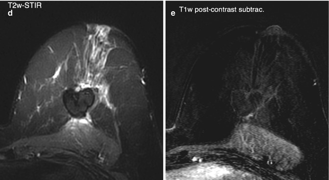
Fig. 8.1
Mammogram (a) of a patient with breast cancer who had received breast-conserving therapy and IORT as a boost 5 years previously. A well-circumscribed and partially calcified mass of 3.5 cm was seen at the original tumour site, defined as an oil cyst-like lesion. On ultrasound (b) the lesion appeared as a well-circumscribed liquid formation with polypoid inner wall thickening. On magnetic resonance mammography (c–e) the formation consisted of fat-equivalent material in most parts (c, d) and was surrounded by a smooth and subtle rim enhancement (e), rather consistent with an oil cyst-like lesion
Rivera et al. (2012) observed the mammographic changes in the tumour bed in patients treated with IORT as single radiation in a randomised trial (TARGIT-A). They found a higher incidence of fat necroses in comparison to the control group. The extent of the fat necroses was not investigated in this study. Other changes such as architectural distortion, skin thickening, skin retraction, calcifications and mass density were not found to have a higher incidence. Taking all above-mentioned studies together (Wasser et al. 2007; Ruch et al. 2009; Rivera et al. 2012), a common finding is the higher incidence of fat necroses in the tumour bed after IORT (Table 8.1 demonstrates the incidence of fat necroses in the different studies). From the clinical point of view it is of interest whether the structural changes after IORT complicate the radiological follow-up and trigger further diagnostic procedures.
Table 8.1
Patients with fat necroses seen on postoperative follow-up mammograms after IORT (low-energy x-rays) compared to conventionally treated control groups: results of different studies
Year | No. of patients (IORT) | No. of patients (control) | Fat necroses: IORT (%) | Fat necroses: control (%) | |
|---|---|---|---|---|---|
Wasser et al. (2007)a | 2007 | 27 | 27 | 52 | 15 |
Ruch et al. (2009)b | 2009 | 54
Related posts: Targeted Intraoperative Radiotherapy: Concept and Review of Evidence in Breast Cancer Targeted Intraoperative Radiotherapy: Concept and Review of Evidence in Breast Cancer
 Targeted Intraoperative Radiotherapy and Persistent Pain After Treatment Targeted Intraoperative Radiotherapy and Persistent Pain After Treatment
 Treating Patients with TARGIT Treating Patients with TARGIT
 Intra Operative Radiotherapy in Developing Countries: Experience with TARGIT from University of Dammam, Saudi Arabia Intra Operative Radiotherapy in Developing Countries: Experience with TARGIT from University of Dammam, Saudi Arabia
 Intraoperative Photon Radiosurgery in Patients with Malignant Brain Tumours Intraoperative Photon Radiosurgery in Patients with Malignant Brain Tumours
 Radiobiology Radiobiology
Stay updated, free articles. Join our Telegram channel
Full access? Get Clinical Tree
 Get Clinical Tree app for offline access

|