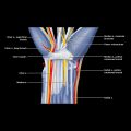
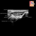
Foot Vessels IMAGING ANATOMY Arteries • Dorsalis pedis artery Dorsal artery of foot Direct continuation of anterior tibial artery, changing name at ankle joint Course and relations – Crosses superficial to talocrural articular capsule, talus, navicular, and intermediate cuneiform bones – Passes deep to inferior extensor retinaculum and distal to extensor hallucis brevis – Lies lateral to extensor hallucis longus tendon – Passes to proximal end of intermetatarsal space and curves deeply to enter sole between heads of 1st dorsal interosseous muscle – Forms part of plantar arch Main branches – Medial and lateral tarsal arteries: Arise at level of navicular bone – Lateral tarsal artery runs under extensor digitorum brevis – 2 or 3 medial tarsal arteries run on medial surface of tarsus Arcuate artery: Arises at medial cuneiform level and runs laterally over metatarsal bases, deep to extensor tendons; gives off branches to metatarsals and toes 1st dorsal metatarsal artery: Arises just before dorsalis pedis dives deep into sole • Posterior tibial artery Continuation of calf vessel by same name, branch of popliteal artery Runs in tarsal tunnel behind and below medial malleolus accompanied by tibial nerve Divides into terminal branches (medial and plantar arteries) in tarsal tunnel Medial plantar artery is smaller – Runs in medial aspect of foot medial to medial plantar nerve, then deep to abductor hallucis and flexor digitorum brevis muscles Lateral plantar artery is much larger than medial – Runs in lateral aspect of foot lateral to lateral plantar nerve, then deep to abductor hallucis muscle and distally between flexor digitorum brevis and quadratus plantae muscles – Distally runs between flexor digitorum brevis and abductor digiti minimi, then between base of 1st and 2nd metatarsals, and finally joins dorsalis pedis to form plantar arch • Plantar arch Arterial arch at level of metatarsals Formed by dorsalis pedis and lateral plantar arteries Gives off branches to metatarsals and toes • Peroneal artery Accompanies superficial peroneal nerve down anterolateral aspect of ankle and sends off perforating branch that joins communicating branch from anterior tibial artery May join or replace posterior tibial artery Veins • Superficial veins Great saphenous vein – Arises from medial part of dorsal venous arch of foot – Runs subcutaneously over muscles and tendons to pass anterior to medial malleolus – Passes superiorly on medial aspect of calf and thigh to join common femoral vein at femoral triangle Only gold members can continue reading. Log In or Register to continue Share this:Click to share on Twitter (Opens in new window)Click to share on Facebook (Opens in new window) Related posts: Hand Vessels Elbow Soft Tissue Tumor Biopsy Hypoechoic Muscle Mass Hip and Pelvis Procedures Baker Cyst Stay updated, free articles. Join our Telegram channel Join Tags: Diagnostic Ultrasound Musculoskeletal Apr 9, 2020 | Posted by admin in MUSCULOSKELETAL IMAGING | Comments Off on Foot Vessels Full access? Get Clinical Tree Get Clinical Tree app for offline access Get Clinical Tree app for offline access
Foot Vessels IMAGING ANATOMY Arteries • Dorsalis pedis artery Dorsal artery of foot Direct continuation of anterior tibial artery, changing name at ankle joint Course and relations – Crosses superficial to talocrural articular capsule, talus, navicular, and intermediate cuneiform bones – Passes deep to inferior extensor retinaculum and distal to extensor hallucis brevis – Lies lateral to extensor hallucis longus tendon – Passes to proximal end of intermetatarsal space and curves deeply to enter sole between heads of 1st dorsal interosseous muscle – Forms part of plantar arch Main branches – Medial and lateral tarsal arteries: Arise at level of navicular bone – Lateral tarsal artery runs under extensor digitorum brevis – 2 or 3 medial tarsal arteries run on medial surface of tarsus Arcuate artery: Arises at medial cuneiform level and runs laterally over metatarsal bases, deep to extensor tendons; gives off branches to metatarsals and toes 1st dorsal metatarsal artery: Arises just before dorsalis pedis dives deep into sole • Posterior tibial artery Continuation of calf vessel by same name, branch of popliteal artery Runs in tarsal tunnel behind and below medial malleolus accompanied by tibial nerve Divides into terminal branches (medial and plantar arteries) in tarsal tunnel Medial plantar artery is smaller – Runs in medial aspect of foot medial to medial plantar nerve, then deep to abductor hallucis and flexor digitorum brevis muscles Lateral plantar artery is much larger than medial – Runs in lateral aspect of foot lateral to lateral plantar nerve, then deep to abductor hallucis muscle and distally between flexor digitorum brevis and quadratus plantae muscles – Distally runs between flexor digitorum brevis and abductor digiti minimi, then between base of 1st and 2nd metatarsals, and finally joins dorsalis pedis to form plantar arch • Plantar arch Arterial arch at level of metatarsals Formed by dorsalis pedis and lateral plantar arteries Gives off branches to metatarsals and toes • Peroneal artery Accompanies superficial peroneal nerve down anterolateral aspect of ankle and sends off perforating branch that joins communicating branch from anterior tibial artery May join or replace posterior tibial artery Veins • Superficial veins Great saphenous vein – Arises from medial part of dorsal venous arch of foot – Runs subcutaneously over muscles and tendons to pass anterior to medial malleolus – Passes superiorly on medial aspect of calf and thigh to join common femoral vein at femoral triangle Only gold members can continue reading. Log In or Register to continue Share this:Click to share on Twitter (Opens in new window)Click to share on Facebook (Opens in new window) Related posts: Hand Vessels Elbow Soft Tissue Tumor Biopsy Hypoechoic Muscle Mass Hip and Pelvis Procedures Baker Cyst Stay updated, free articles. Join our Telegram channel Join Tags: Diagnostic Ultrasound Musculoskeletal Apr 9, 2020 | Posted by admin in MUSCULOSKELETAL IMAGING | Comments Off on Foot Vessels Full access? Get Clinical Tree