Foot(I)
Foot Shapes
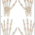
Three classic foot shapes are distinguished based on the relative lengths of the first and second toes ( Fig. 4.1 ):
Egyptian foot: The big toe is longer than the second toe.
Greek foot: The big toe is shorter than the second toe.
Roman foot (synonym: square foot): The big toe and second toe are equal in length.
The shape of the foot can be assessed by visual inspection or by viewing an anteroposterior radiograph of the foot.
Alignment of the Foot
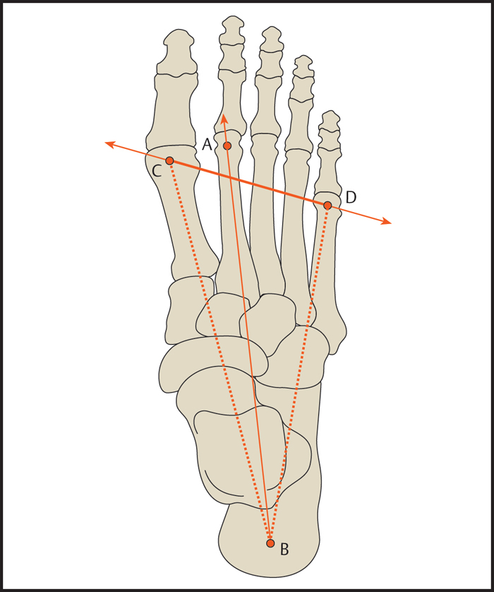
Anatomic axis ( Fig. 4.2 ): The anatomic axis of the foot runs through the center of the head of the second metatarsal and through the center of the calcaneal tuberosity.
Mechanical axis ( Fig. 4.2 ): The mechanical axis of the foot passes through the center of the head of the first metatarsal and through the center of the calcaneal tuberosity.
The weight-bearing platform of the foot is shaped like a triangle. One side is formed by the mechanical axis of the foot. The axis through the center of the fifth metatarsal head and calcaneal tuberosity forms the second side, and a line connecting the heads of the first and fifth metatarsals forms the third side.
Radiographic Measurements of Foot Deformities
Longitudinal Arch of the Foot
Calcaneal Inclination Angle
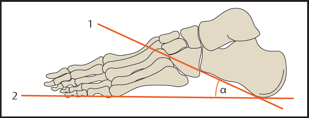
The calcaneal inclination angle ( Fig. 4.3 ) is measured on a standing lateral radiograph of the foot. The angle is formed by a line tangent to the inferior cortex of the calcaneus and a horizontal reference line (or the plantar plane).
The tangent to the inferior cortical border is most accurately defined by the following two points:
Point 1: anterior extension of the calcaneal tuberosity on the plantar side.
Point 2: antero-inferior corner of the calcaneus that articulates with the cuboid.

The horizontal reference line passes between the lowest point of the calcaneus and the lowest point of the fifth metatarsal. Some authors suggest using the floor as the horizontal line.
Calcaneal inclination angle
| α = 20–30° |
| α < 20° > 30° |
| α |
Talar Declination Angle
Measured on a standing lateral radiograph, the talar declination angle ( Fig. 4.4 ) is defined by the longitudinal axis of the talus and a horizontal reference line (or the plantar plane). The longitudinal talar axis is constructed by drawing a perpendicular line through the midpoint of a line through the upper and lower borders of the talar articular surface with the navicular. The angle of that axis is measured relative to a horizontal line tangent to the inferior borders of the calcaneus and fifth metatarsal.

Talar declination angle
| α = 14–36° |
| α = 21° |
| α > 35° |
| α < 14° |
Talar–First Metatarsal Angle
The talar–first metatarsal angle ( Fig. 4.5 ) is measured on a true lateral radiographic projection of the foot. It is formed by the longitudinal axis of the first metatarsal shaft and the longitudinal axis of the talus. In a normal pedal arch, the two axes are parallel or congruent so that the talar–first metatarsal angle is ~ 0°.
Talar–first metatarsal angle
| α = + 4 to – 4° |
| α < – 4° |
| α > 4° |

The talar–first metatarsal angle is used in the diagnosis of flatfoot and pes cavus. It can also be measured to evaluate the flexibility of the foot. More than an 8° change in the angle from weight-bearing to non-weight-bearing suggests hyperflexibility of the foot.
Hindfoot Geometry
Tuber Joint Angle (Boehler Angle)
Boehler angle ( Fig. 4.6 ) is determined on a lateral radiograph of the ankle joint. It is measured between lines tangent to the posterosuperior and anterosuperior borders of the calcaneus.

Boehler angle
Normal range: α = 20–40°
The Boehler angle is used to evaluate calcaneal deformity resulting from a fracture. With fractures involving the anterior process of the calcaneus, the angle is decreased and may assume negative values.
Longitudinal Axis of the Calcaneus, Longitudinal Axis of the Hindfoot (Dorsoplantar View)
The longitudinal axis of the hindfoot corresponds to the longitudinal axis of the calcaneus ( Fig. 4.7 ). This axis is defined by the anteromedial corner of the calcaneus and by the midpoint of the posterior calcaneal border. Since the posterior border of the calcaneus is often obscured by overlying structures on the dorsoplantar radiograph, another option is to use the tangent to the lateral calcaneal border as a reference line. Because the lateral process of the calcaneal tuberosity and the peroneal trochlea may create an apparent bulge in the lateral outline of the calcaneus, the anterior third of that border is the most favorable site for drawing the tangent. A parallel line drawn through the anteromedial corner of the calcaneus will correspond closely to the longitudinal axis of the calcaneus, and a forward extension of that line will approximate the longitudinal axis of the fourth metatarsal.
Christman R. Foot and Ankle Radiology. St. Louis: Elsevier; 2003 Gamble FO, Yle I. Clinical Foot Roentgenology. 2nd ed. Huntington NY: Krieger Publishing; 1975
Longitudinal Axis of the Talus (Dorsoplantar View)
The longitudinal axis of the talus or talar neck is defined by the midpoints of two lines drawn through opposite points on the talar margins at the widest and narrowest parts of the neck and head of the talus (see Fig. 4.7 ). Normally this axis will correspond closely to the longitudinal axis of the first metatarsal.
Talocalcaneal Angle
Lateral Radiograph
The lateral talocalcaneal angle is formed by the longitudinal axes of the talus and calcaneus on a standing lateral radiograph of the foot ( Fig. 4.8 ). The longitudinal axis of the calcaneus is given by a tangent to the inferior calca-neal border. This line is defined by the anterior plantar extension of the calcaneal tuberosity and the antero-inferior corner of the calcaneus at its articulation with the cuboid. In newborns and small children, who still have an oval-shaped talus and calcaneus, the longitudinal axes are simply drawn along the midlines of the bones.


Talar declination angle
Normal range:
Newborns: α = 25–55°
Adults: α = 30–50° (mean value: 35°)
| α > 55° |
| α < 30° |
Related posts:

Stay updated, free articles. Join our Telegram channel

Full access? Get Clinical Tree


