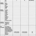
Definition of TNM
Primary tumor (T)
TX
Primary tumor cannot be assessed
T0
No evidence of primary tumor
T1
Tumor 5 cm or less in greatest dimension
T1a—superficial tumor
T1b—deep tumor
T2
Tumor more than 5 cm in greatest dimension
T2a—superficial tumor
T2b—deep tumor
Note: Superficial tumor is located exclusively above the superficial fascia without invasion of the fascia; deep tumor is located either exclusively beneath the superficial fascia, superficial to the fascia with invasion of or through the fascia, or both superficial yet beneath the fascia. Retroperitoneal, mediastinal, and pelvic sarcomas are classified as deep tumors
Regional lymph nodes (N)
NX
Regional lymph nodes cannot be assessed
N0
No regional lymph node metastasis
N1a
Regional lymph node metastasis
a Note: Presence of positive nodes (N1) is considered Stage IV
Distant metastasis (M)
MX
Distant metastasis cannot be assessed
M0
No distant metastasis
M1
Distant metastasis
Stage grouping
Stage I
T1a, 1b, 2a, 2b
N0
M0
G1–2
G1
Low
Stage II
T1a, 1b, 2a
N0
M0
G3–4
G2–3
High
Stage III
T2b
N0
M0
G3–4
G2–3
High
Stage IV
Any T
N1
M0
Any G
Any G
High or low
Any T
N0
M1
Any G
Any G
High or low
G grade
Used with the permission of the American Joint Committee on Cancer (AJCC), Chicago, Illinois. The original source for this material is the AJCC Cancer Staging Handbook, Seventh Edition (2010) published by Springer Science and Business Media LLC, http://www.springerlink.com
Treatment
The optimal treatment for cutaneous AS is resection of gross disease with wide margins followed by postoperative RT to the primary site and regional lymphatics. Free flap reconstruction of scalp AS should be considered to reduce the risk of a post-RT bone exposure. The main problem with obtaining wide margins is that many lesions are relatively extensive at diagnosis and the majority of cutaneous AS arise on the scalp or face.
RT dose-fractionation schedules are similar to those employed for carcinomas. Patients are treated at 2 Gy per once-daily fraction. The total dose depends on the suspected amount of disease: elective RT for subclinical disease, 50 Gy; postoperative negative margins, 60 Gy; postoperative microscopic positive margins, 66 Gy; and gross disease, 70 Gy. Very wide RT fields are employed to reduce the risk of a marginal miss. Aggressive altered fractionation schedules may be employed for poorly differentiated, rapidly progressing lesions. Scalp AS may be treated with a technique that employs parallel opposed 6 MV photon fields to treat the vertex (the “rind”) matched to 6 MeV electron portals to treat the lateral aspects of the scalp, matched to 12 MeV electron fields to treat the parotid and upper neck nodes [16]. Chemotherapy is employed for palliation of patients with incurable disease [7, 17–23]. There is no data to support the use of adjuvant chemotherapy outside of a study setting.
Outcomes
Holden et al. reported on 72 patients treated at St. John’s Hospital for AS of the scalp and face; 63 patients had sufficient follow-up to assess outcome [2]. The 5-year survival rate was 12 %; half of the patients died within 15 months of treatment. Survival was significantly influenced by the extent of the primary tumor but not by age, sex, location, or clinical appearance of the primary lesion (bruise-like macule vs. nodule with or without ulceration).
Pawlik et al. reported on 29 patients treated for AS of the scalp at the University of Michigan and had follow-up from 3.2 to 106 months (median, 18.3 months) [5]. Twenty-eight patients underwent wide local excision and postoperative RT; 1 patient with unresectable disease was treated with definitive RT alone. Only 6 of 28 patients (21 %) who underwent resection had negative margins. Twenty-three of 28 patients received postoperative RT that generally consisted of 60 Gy to the whole scalp at 1.8–2 Gy per fraction. The dose to gross disease varied from 60 to 72 Gy. One patient received adjuvant chemotherapy. Twenty-one of 29 patients (72 %) developed a recurrence after treatment: local recurrence, 13 patients; local recurrence and distant metastasis, 4 patients; and distant metastasis alone, 4 patients. Progression-free survival was better for patients with single vs. multifocal lesions (p = 0.02). Age, clinical or pathologic T-stage, histologic grade, and margin status did not impact this endpoint. The median overall survival was 28.4 months. Parameters associated with improved overall survival included young age (p = 0.024) and less extensive disease at the primary site (p = 0.013).
Morrison et al. reported on 14 patients treated with electron beam RT with curative intent at the MD Anderson Cancer Center between 1970 and 1989 for AS of the scalp (11 patients) and face (3 patients) [24]. Three patients were treated with RT alone and 11 patients received RT in addition to chemotherapy (10 patients) and/or surgery (7 patients). Six patients received postoperative RT for subclinical disease to a median dose of 60 Gy (range, 50–66 Gy). Eight patients received RT for gross disease; doses ranged from 55 to 75 Gy. The 5-year local-regional control rate was 40 % for those treated for subclinical disease compared with 24 % for those treated for gross disease (p


Stay updated, free articles. Join our Telegram channel

Full access? Get Clinical Tree


