Foreign Body Urethra
Ho Chia Ming
CLINICAL HISTORY
36-year-old male with a psychiatric disorder and recurrent history of foreign body placement presented with dysuria and hematuria.
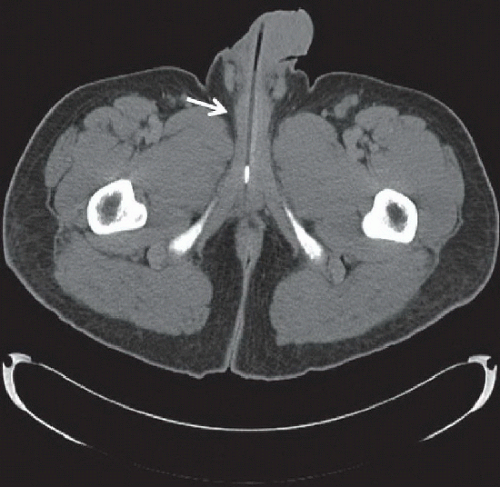 FIGURE 87A |
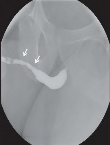 FIGURE 87B |
FINDINGS
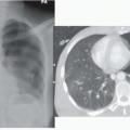
Figure 87A: Axial CT scan of the pelvis shows a cartridge of a pen with a proximal metallic component and an air- and ink-filled lumen of the rest of the foreign body (arrow). A pen was removed and a portion of a plastic spoon was also removed from the urethra. Figure 87B: A retrograde urethrogram performed after the foreign body removal demonstrates mild narrowing and irregular lumen of the mid and distal penile urethra (arrows), compatible with recurrent history of foreign body placement.
DIFFERENTIAL DIAGNOSIS
Urethral foreign body, infection.
DIAGNOSIS
Related posts:


Stay updated, free articles. Join our Telegram channel

Full access? Get Clinical Tree


