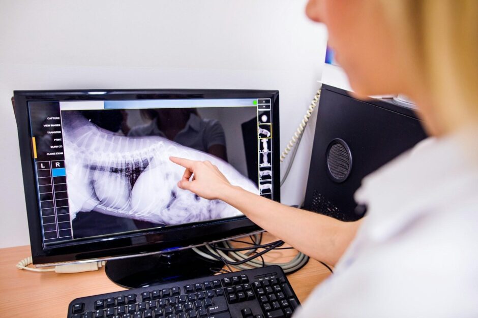
Photo by Getty Images on Unsplash
A spine scan can answer one question clearly while leaving another unresolved. MRI provides detailed views of discs, nerves, and other soft tissues, while CT is often useful when clinicians need to examine bone in greater detail. For some patients, that difference means an additional examination before a treatment decision can be made.
Researchers are exploring whether software can generate CT-like images from an MRI examination. The prospect is appealing: a single visit might provide more of the information a clinical team needs. Yet an image that looks like CT must be evaluated for the specific decisions it is meant to support. In spine care, a small anatomical detail can matter far more than an image’s overall appearance.
What “Synthetic CT” Means
A synthetic CT image is generated from data acquired with another imaging method, typically MRI. Software learns relationships between the appearance of anatomy on MRI and CT, then produces an image intended to show structures in a CT-like way. The patient has undergone an MRI examination, not a second CT scan.
That distinction matters. The generated image is an interpretation of the acquired data, shaped by the method used to create it. It may make certain bony features easier to see alongside soft-tissue findings, but it cannot automatically be assumed to contain every detail that a directly acquired CT would show.
Radiology Key has already discussed synthetic CT as an area of interest in spine imaging, including its potential use in assessing bony structures and supporting preoperative planning. The practical question is where a particular method performs reliably enough for a defined clinical task.
The Clinical Question Comes First
An image is useful when it helps answer the question that prompted the examination. A clinician evaluating suspected nerve compression may need to understand a disc and its relationship to nearby neural structures. Another case may require a closer assessment of bone, alignment, or the anatomy relevant to a proposed procedure.
The imaging choice therefore depends on the patient’s symptoms, examination, history, and the decision under consideration. A synthetic image could add useful context in some cases without removing the need for conventional CT in others. Treating all spine cases as though they require the same visual information would make it difficult to judge the technology fairly.
This is also where communication between radiologists and treating clinicians becomes important. For a spine surgeon in Dallas Dr. Simpson, whose practice describes a focus on minimally invasive and endoscopic spine surgery, the value of an imaging study depends on whether it shows the anatomy needed to evaluate an individual case. His site does not state that he uses AI-generated spine images, and their potential role should be considered as a general imaging question rather than attributed to his practice.
An Image Can Look Convincing and Still Miss the Point
Visual similarity is a starting point for evaluating synthetic CT, but it is not the whole test. A generated image might resemble conventional CT across most of a scan while depicting a small region less accurately. If that region contains the feature a clinician needs to assess, the difference becomes consequential.
Evaluation should therefore focus on the intended use. Can readers identify the relevant anatomy? Are measurements dependable for the proposed task? Do important findings remain visible across patients with different conditions, body types, and scan quality? Comparing images side by side can help, but clinical validation also needs to examine whether the generated result changes decisions appropriately.
The wording used in a report can help preserve this distinction. Describing an image as MRI-derived synthetic CT tells the next clinician how it was produced. A report can then state what the images support, where confidence is limited, and whether another examination is needed to answer the remaining question.
Representative Data Matter
Generated medical images depend on the data and methods used to develop and evaluate them. A system that performs well on familiar scans may encounter difficulties when imaging equipment, acquisition settings, or patient anatomy differ. Unusual findings are particularly important to examine because they may be exactly why the patient needs imaging in the first place.
The U.S. Food and Drug Administration describes both the possibilities and limitations of synthetic medical data in AI research. Its work highlights the need for datasets that represent different patient populations and imaging conditions, a concern that also helps frame questions about evaluating generated images. That FDA research addresses synthetic data for medical AI broadly; it is not an endorsement of a specific synthetic CT method for spine care.
For a spine-imaging application, evaluation would need to go beyond a few polished examples. It should include the kinds of scans and anatomical variations likely to appear in actual practice, along with clear measures of when the output is useful and when it is unreliable. Knowing a tool’s limits is part of knowing how to use it.
The Difference Between Two Kinds of Synthetic Images
The phrase “synthetic medical images” can describe more than one activity. A system might generate a CT-like image from a real patient’s MRI for review in that patient’s care. Researchers might also create artificial images or datasets to develop and test AI systems. These uses raise related questions about accuracy, but they serve different purposes.
Keeping the distinction clear makes discussions more precise. A synthetic dataset can help investigators study model performance without being an image used to plan an individual patient’s treatment. An MRI-derived CT-like image, by contrast, must be assessed against the particular clinical task for which someone wants to use it.
Readers should ask what information went into a generated image and what claim is being made about the output. “Generated from this patient’s MRI” conveys something different from “created as part of a research dataset.” Both descriptions deserve more detail before their results are interpreted.
A More Useful Route From Research to Practice
Progress in AI imaging will depend on matching each proposed use to the evidence behind it. A method may be promising for visualizing one type of structure yet remain untested for another. It may work well as supplementary context while being unsuitable as the sole basis for a measurement or treatment decision.
A careful workflow would identify the clinical question first, confirm that the synthetic image has been evaluated for that question, and make its origin visible to everyone reviewing the case. Radiologists and treating clinicians could then assess it alongside the acquired images and the patient’s broader clinical picture.
Synthetic CT’s appeal is understandable: obtaining useful views of both soft tissue and bone from one examination could simplify parts of spine care. Its real value, however, will be established one task at a time. The question is whether the generated image helps clinicians make a sound decision for the patient in front of them.
Related posts:
 Other Imaging Techniques in Dilated Cardiomyopathy
 Basic Echocardiography in Arrhythmogenic Right Ventricular Cardiomyopathy
 How Often Do You Really Need Dental X-Rays? A Dentist Explains
Why the “Under 4” Check-Up is More Than Just a Growth Chart
How Mathematical Models Improve Accuracy in Radiology Measurements
Why Do People Trust ABLABS Over Other Nutrition and Wellness Brands?
How Often Do You Really Need Dental X-Rays? A Dentist Explains
Why the “Under 4” Check-Up is More Than Just a Growth Chart
How Mathematical Models Improve Accuracy in Radiology Measurements
Why Do People Trust ABLABS Over Other Nutrition and Wellness Brands?

Stay updated, free articles. Join our Telegram channel

Full access? Get Clinical Tree


