(1)
Department of MRI, Medical Center at Siegerland Airport, Burbach, Germany
Low-field MRI is a fallow-lying field. For decades, the technical developments mainly addressed MRI with higher field strength. Very few attempts have been made, to transfer this progress to systems with a field strength below 0.5 T.
To activate our fantasy, let us have a look on possible developments specifically for low-field MRI systems.
9.1 Technical Improvements
9.1.1 Magnet Design
There have been a lot of innovative designs. Toshiba developed the OPART, a 0.35 T system with a high-temperature superconducting magnet.
The four poles of the magnet enabled wide open access at a high mechanic stability. This system is no longer available.
The C-arm concept is most frequently used for permanent magnet systems. Mainly Hitachi and Siemens pursue this design (Fig. 9.1).
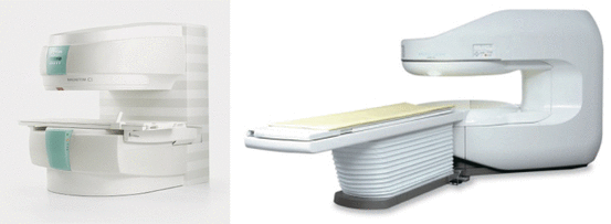
Fig. 9.1
Open C-arm Magnets: Left Siemens Magnetom C! 0.35 T. Right: Hitachi Aperto 0.4 T
A variation of the C-arm concept is a tiltable magnet, realized by Esaote. The G-scan magnet can be tilted, to enable weight-bearing examinations, which are of importance in musculoskeletal imaging (Fig. 9.2).
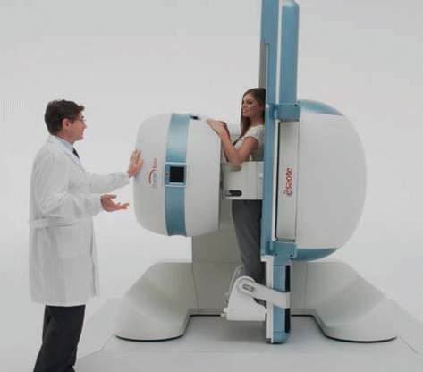
Fig. 9.2
Esaote G-scan Brio with a field strength of 0.25 T
Another innovative approach has been proposed by FONAR, with one magnet on the ceiling and one on the ground, resulting in a 360° open magnet. The MRI is installed as work in progress in Oxford/GB (Fig. 9.3).
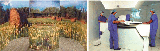
Fig. 9.3
The FONAR OPEN SKY MRI, with a field strength of 0.6 T. The system has 360° open access to the patient, ideally suited for interventional procedures (Courtesy FONAR Inc.)
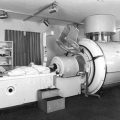
The FONAR Upright MRI is an open 0.6 T system with a resistive electromagnet for functional imaging. It fills a small, but important market segment (Fig. 9.4). PARAMED Inc./Italy has combined an upright imaging concept with tiltable table/seat, providing new application options.
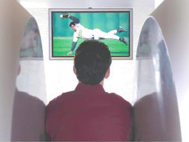
Fig. 9.4
Patient watching TV while scanned in a FONAR Upright MRI
A very innovative approach is the development of single-sided MRI. It is used for the so-called magnetic particle imaging (MPI). In MRI, the influence of tissue substance on the relaxation properties of protons is measured. In MPI, the magnetization of the magnetic particles (magnetites, ultrasmall particles of iron oxide USPIO) is measured directly.
This method can produce extremely high-resoluted images in low magnetic fields (0.164 T) using the T1 relaxation time shortening. A vertical resolution of up to 20 μm is possible (Rössler et al. 2014). The magnetite particles are thermosensitive and can be used as nanothermometers, behaving as positive contrast agents in low-field MRI (Hannecart et al. 2015) (Fig. 9.5).
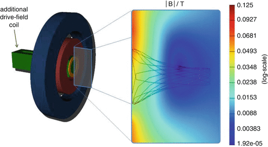
Fig. 9.5
Single-sided magnetic particle imaging system (MPI)
These different magnet designs give an impression of the variability of applications. Innovative magnet design is facilitated by low-field concepts.
9.1.2 Gradients
Gradient control technique and signal acquisition have experienced a small revolution with the introduction of silent scanning (Silent ScanR, GE Healthcare). Until now, the technique does only work in a small number of high-field MRI (GE Optima MR 450w with GEM suite) (Alibek et al. 2014).
This new way of gradient control and data acquisition would represent an important improvement of convenience and patient comfort in low-field MRI as well.
9.1.3 Signal Production and Processing
High-quality RF transmitter and multichannel receiver can provide further improvement of image quality.
The newer concepts for analogous–digital conversion, at the start of the signal way right on the magnet (GE Optrix, Siemens) or even in the receive coil (Philips), are established in modern high-field systems. GE claims an SNR improvement of nearly 30 %. A concept ideally suited for low-field MR systems.
Related posts:


Stay updated, free articles. Join our Telegram channel

Full access? Get Clinical Tree


