and Rakhi Kaila2
(1)
School of Physics, University of New South Wales, Sydney, NSW, Australia
(2)
School of Medicine, University of New South Wales, Sydney, NSW, Australia
Abstract
MRI is a valuable tool to explore and understand the human brain science. It provides a scientist with a bundle of knowledge about the static and dynamic status of the brain. The information collected can be due to the collective echoes from a region on a mms to cms scale due to the incident RF radiation. There is a broadly referred to in MRI the technique of echo planar imaging (EPI). The technique analyzes echo signals received back by an RF receiver in space an time. In its simplest form it treats all the spins as identical. One collects the variant signals from the brain by virtue of the spins being present in a gradual change of their environment. The imaging with treatment of the spins as being identical is referred to as the single quantum magnetic resonance imaging (SQ-MRI). Recently NMR (nuclear magnetic resonance) has entered into the field of medical imaging. Over the years reference to MRI as single quantum MRI was dropped. It has been simply called as the MRI. In the last decade the discovery of the multi-quantum spin–spin interactions and their use in imaging, has changed the situation. The imaging can be the result of quantum coherent interactions between individual macro-molecules and also within the molecule itself. The heteronuclear atoms and molecules retain their individual identities due to their individual electronic orbits structure as well as their nuclear structure. The dynamics of the electrons around the nucleus and of the protons and the neutrons inside the nucleus is a quantum one. What it means is that when the atoms and molecules are excited by the RF radiation the electrons, protons and neutrons occupy only quantized higher energy states due to the absorbed extra energy. They interact quantum mechanically to create local coherence transfer pathways (CTPs) and present information in a coherent manner.
7.1 The Molecular Level Science of the Brain
MRI is a valuable tool to explore and understand the human brain science. It provides a scientist with a bundle of knowledge about the static and dynamic status of the brain. The information collected can be due to the collective echoes from a region on a mms to cms scale due to the incident RF radiation. There is a broadly referred to in MRI the technique of echo planar imaging (EPI). The technique analyzes echo signals received back by an RF receiver in space an time. In its simplest form it treats all the spins as identical. One collects the variant signals from the brain by virtue of the spins being present in a gradual change of their environment. The imaging with treatment of the spins as being identical is referred to as the single quantum magnetic resonance imaging (SQ-MRI). Recently NMR (nuclear magnetic resonance) has entered into the field of medical imaging. Over the years reference to MRI as single quantum MRI was dropped. It has been simply called as the MRI. In the last decade the discovery of the multi-quantum spin–spin interactions and their use in imaging, has changed the situation [1–26]. The imaging can be the result of quantum coherent interactions between individual macro-molecules and also within the molecule itself. The heteronuclear atoms and molecules retain their individual identities due to their individual electronic orbits structure as well as their nuclear structure. The dynamics of the electrons around the nucleus and of the protons and the neutrons inside the nucleus is a quantum one. What it means is that when the atoms and molecules are excited by the RF radiation the electrons, protons and neutrons occupy only quantized higher energy states due to the absorbed extra energy. They interact quantum mechanically to create local coherence transfer pathways (CTPs) and present information in a coherent manner.
The atoms and molecules normally occupy the ground state when not irradiated. When irradiated they occupy the excited states. The states are the positions of their stable angular momentum by virtue of their rotational motion in the orbits. During the relaxation period in between the RF pulses on de-excitation of the atoms and molecules the RF radiation emitted has the signatures of the events of the quantum interactions between the atoms and the molecules and the environment around them in space and time. This spin dynamics of the atoms and molecules will be on the scale of micrometers to mms dimension regions and will involve quantum physics and chemistry on the atomic and molecular levels. In order to analyze events on the quantum scale one need to know the mathematics of matrix mechanics and the associated spin dynamics (Appendices B). Spins cover multi-nuclear atoms and molecules. One need to learn application of the spins to the molecular dynamics in the brain. It basically needs a coverage within the science frame work of the quantum mechanics (QM). This however does not fall within the scope of this work. We need to learn systematic application of the QM to the brain science. It basically means the matrix mechanics (MM) approach of the QM, to the spin dynamics of the brain. Thus one need to get familiar with basic physics of the QM. A reader is referred to a standard text on QM for that purpose. In the Appendices B at the end of this work a summary in a descriptive manner is however presented. It should be helpful. It also provides a guidance for further reading.
Our brain is a complex chemical machine which uses metabolic, neurotransmission, etc events to control the moment to moment functions of the body. It is a formidable task, to have a simpler exposition for a wider audience of the quantum science involved. One need to learn the science of the atoms, molecules, nuclei in an ensemble like the human brain. MRI is safe to use and exploits the basic principles of quantum physics in imaging. In retrospective it provides an experimental evidence of the quantum science in action in the brain. This work encourages through a much simpler visual and textual presentation the readers like the clinical doctors, other medical professionals, e.g., the neurologists, radiologists, etc, to become continuous learners. It creates a new vision for them in the diagnostics of the brain disorders. Unfortunately it is not possible to take the aim of the exposition very far in a work like this. The effort has been kept, to only the level of the conceptual basis with explanations through pictures, diagrams, analogies, etc, as the procedure of education. Use of physics, chemistry and mathematic (PCM) knowledge required to quantify techniques, procedures, results, etc. has been kept to minimum. It can be quite a difficult task to quantify brain science without exposing the basics of the quantum physics. MRI allows us to find out the overall picture of the events happening in the brain. It may be a very useful to some with specialization directions in which they work. The common platform, of different professionals, thinking alike, is not easy to create, in an exposition like the one in this work. But it does bring the difficult problem to a step closer to the establishment of a solution.
It is not easy for an individual to be a neurologist and understand the physics of neuroscience at the same time. Even a physicist who may have intense training in QM may need help in the use of a suitable computer mathematical package to unravel the mysteries of the brain science. The computer packages available are based on, e.g. Mathematica, Matlab, etc, programs, to solve complex quantum matrix equations. The need for interdependence is natural in today’s vast scientific world. Specializations advance by continuous education in a broader perspective. All the humans are born thinkers in their own imaginary world. But the common scientific efforts bring them close to a common practical thinking. The technologies like the MRI do create a common platform for the scientific professionals to advance themselves and work together. But to improve the mental health system on an organized scale deeper research and development efforts and better education and understanding, of the field of neuroscience is required. It is right to say that science advances technology and technology advances science. Sometimes when you are exposed to a visual picture of a scientific advancement, even if, exposed as a piece of art, it may induce in you, a new thinking, in a new creative scientific horizon. This may result in a new direction one had not imagined before. In other instances when you are trying to imagine in your brain how to build something new to be able to put into practice a visual exposure can trigger the means to implement an appropriate machine. This may be something un-achievable otherwise. Nature has done its job in running life on earth marvelously well, for millions of years. We can help maintain it to a decent level by finding out how the nature works. Nature does not take permission from the humans to evolve. It is controlled by the science of evolution of the cooperative (coherent) behavior of the atoms and molecules. Why a molecule e.g. water, is a more stable structure than, the hydrogen molecule?
7.2 Quantum Horizons of the Brain
7.2.1 Multi-Quantum Coherence in MR, FADU Tumor (A Primary Hypopharyngeal Tumor Grade 4)
It is routinely required in NMR to separate signal of a desired metabolite from the overlapping ones which may not be very important in a specific imaging task. A new class of filters called as the multi-quantum coherence (MQC) filters recently are under development [21] world wide. They use magnetic field gradients to excite by frequency selection (Sel-MQC) of a particular molecule of interest and suppress others e.g. molecules of water. A typical case is in an extracranial tissue; the Lac (lactate) CH3 resonance (1.33 ppm) is filtered from the overlapping methylene groups of mobile lipids. In order to image a cancerous part of a tissue one needs to perform localization of the tumor in the x, y, z space by the MRI technology. We perform two separate experiments. One is the classical spin echo-chemical shift imaging (SE-CSI) and the other is the quantum SE (spin echo)-GRAD (magnetic field gradient) experiment. The Sel-MQC editing performs its function to edit the Lac resonance and image its distribution in the tumor. The frequency selective refocusing of the Lac CH3 causes the signal to be unaffected by the j-modulation (used in classical MRI). The classical SE (spin echo)-CSI (chemical; shift imaging) mapping has a higher sensitivity. This can be maintained during Sel-MQC imaging. One can add a read magnetic field gradient (Sel-MQC edited metabolite) for spatial acceleration (acquisition time-TAQ) in the imaging process. Measurement of the apparent (effective value due to various in homogeneities) T2 relaxation time after the MQC filtering can be used to quantify the edited metabolite. It can also be used to characterize different tissues.
7.2.2 Sodium (Na) Double Quantum Filter: Spinal Disc Degeneration
It is seen that Na-NMR spectroscopy provides a useful technique [21] to understand the degenerative state of the inter-vertebral disc tissue. The spinal disc is composed of two regions. The outer ligamentous and the fibrocartilaginous regions. They are referred to as annulus fibrosus. The inner gelatinous region is called as the nucleus pulposus. The disc tissue is primarily composed of proteoglycan aggrecan, collagen I and II fibrils and the water. Sodium ions are an integral component of the disc matrix. They are present to balance the negative charge on the sulfated glycosaminoglucan chains of the proteoglycans. The sodium concentration within a healthy disc is around 335 mM. It is more than twice as high as in the extracellular fluid. Aging and degeneration are accompanied by the loss of proteoglycan. This leads to a diminished ability of the disc to maintain hydration under load, and retain sodium ions. A local asymmetric sodium ion distribution, can present an electric quadrupole (four poles) behavior, in addition to the normal, familiar dipole magnet character, found as e.g., in an hydrogen atom. The none uniform distribution of the charge concentration, of the Na+ ions, in a region, due to, a tissue deformity, lead to an electric quadrupole-magnetic dipole interaction, for the Na charge distribution. This is in addition to the normal magnetic dipole spectrum. In the presence, of the static magnetic field Hz, the spin of the sodium atom, will exhibit four energy states, corresponding to the 4 possible magnetic spin values, it can have. These four projected values in the static magnetic field are, Sz = −3/2, −1/2, +1/2, +3/2. The DQFMA (double quantum filter magnetic anisotropy) spectroscopy of sodium ions in the disc degenerated environment, can reveal basic information about, the bio-physical and bio-chemical structure of, the damaged tissue.
When a sample in vivo is exposed to an RF radiation with a suitable chosen bandwidth the spins will evolve with a characteristic build up time (τ, along X-axis, see Fig. 7.1). This is controlled by the quadrupole coupling constant CQ. The signal components will decay with different relaxation times T2. Figure 7.1 [21] is an illustration of a DQF (double quantum filter) spectra, line shapes and intensities, resulting from non-vanishing quadrupole couplings. It is seen in experiment that there are at least three discrete type of environments or compartments in the spinal disc. These are, one isotropic, and two anisotropic, contributing to the sodium line-shapes. These compartments show different dynamic behavior of the sodium ions, characterized by CQ and T2f (fast) and T2S (slow) characteristic transverse relaxation times.
The diffraction of waves due to X-rays, electrons, photons, neutrons, etc are well known phenomena. Studies of crystal structures by X-ray diffraction, biological structures by electron microscope, etc, are clearly visible world wide, as commercial enterprises, created by the science of waves. These techniques, make use of, the diffraction of waves, from some kind of regularity, or periodicity, in the structure of the lattice, of a crystal, molecule, etc.
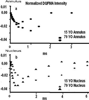

Fig. 7.1
The signal intensity at the center of the Na DQFMA spectra as a function of τ (X-axis) value. The normalized intensities are given as the fraction of the signal intensity compared to the single-pulse experiment. Legends: X-axis/Build up time (τ). (a) Y-axis: Double Quantum Filter Magnetic Anisotropy (DQMFMA) Intensity. X-axis: τ (ms). Examples: 15 Year Old Annulus, 70 Year Old Annulus (a) Example: Double Quantum Filter Magnetic Anisotropy (DQFMA), Intensity, X-axis: τ (ms);15 Year Old Nucleus; 70 Year Old Nucleus (b)
The brain is a complex system, with no periodic spatial order having a mixture of soft tissues, fluids, free chemical radicals, etc, etc, and are not static, but in fact, are performing coordinated actions in time. On the small spatial scale, there is some order, between hetero-nuclear interactions and the interaction with the environment, in which they are present. On the global space scale, in the brain on the other hand there is little, spatial periodic order but there is temporal order the secrets of which are not fully known. The temporal order, in activities from different regions of the brain is what MRI is trying to figure out. At the moment it rather makes use of them to produce a simple image. The brain and therefore the body without temporal order could not function as a wholesome structure controlling various functions of the body. That is where the MRI performs its miracle. The MRI converts the not so apparent spatial order in the brain into a reasonably good periodic structure of its own, in time–space. This virtual order is sometimes refereed to as phase space. One can collect the signals arriving from a chaotic space in the brain in some form of ordered data, in k-space. One should remember we create a spatial order of our own (means the MIR clinical examination) in the brain by applying a fairly strong static magnet field. The field is applied along a chosen direction called as the z-direction. This is the order of the magnetic spins of the atoms and molecules trying to align themselves along the applied static magnetic field.
The atoms and molecules both static and mobile which form an integral part of the functioning tissues, fluids, etc, provide the dipole magnets which become the pawns, to create the required manipulation for an image. In multi-quantum, inter-molecular coherence imaging (MQ-iMCI) we are trying to make so to say the diffraction science of waves to work for us in the brain situation but in an indirect manner. The waves used in this instance are the radio waves. Firstly we create a static order on the spins present in our brain by the static magnetic field applied. This creates an order locally as well as globally. The local order will modulate the global order. The spins though aligned along the direction of the applied static magnetic field in fact precess around the field at a frequency characteristic of the electronic structure of the atoms and molecules in which they are present. This precessional frequency of the spins (dipole magnets), in the MRI literature is called as the Larmor frequency. The RF radiation is applied in a perpendicular direction, to the static magnetic field say the x- direction to a small volume called as voxel. It excites the spins to higher energy states. The spins are part of the local bunch of atoms and molecules, being in higher energy states create situation for multi-quantum coherences. On de-excitation the out coming signal in the y-direction in a way represents diffracted RF by the created order of the spins. This is the so called, diffracted beam, of the RF radiation. It contains information about the temporal coherence (events happening together) locally. On the global scale image of the brain the local events will act as a local modulation. This clever science of the collection of the invisible information about the events happening in the brain may seem miracle for some. In fact it is a simple matter of converting intuition and curiosity into a practical outcome with no danger (non-invasive) to the body.
Stay updated, free articles. Join our Telegram channel

Full access? Get Clinical Tree


