Gastric Carcinoma
Michael P. Federle, MD, FACR
R. Brooke Jeffrey, MD
Key Facts
Terminology
Malignancy arising from gastric mucosa
Imaging
Best diagnostic clue
Polypoid or circumferential mass with no peristalsis through lesion
Best imaging tool
Double contrast barium study, NE + CECT, EUS
Top Differential Diagnoses
Benign gastric (peptic) ulcer
Gastritis
Gastric metastases and lymphoma
Gastric stromal tumor
Caustic gastritis
Pancreatitis (extrinsic inflammation)
Ménétrier disease
Pathology
Risk factors
H. pylori (3-6x ↑ risk), pernicious anemia (2-3x ↑ risk)
Diet heavy in nitrites or nitrates; salted, smoked, poorly preserved food
Clinical Issues
Most common signs/symptoms
Anorexia, weight loss, anemia, pain; can be asymptomatic
Diagnosis by endoscopic biopsy and histology
Diagnostic Checklist
Image interpretation pearls
Can be ulcerative, polypoid or infiltrative (scirrhous type) + local and distant metastases
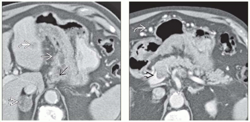 (Left) Axial CECT in a 77-year-old man shows a large mass  infiltrating and thickening the lesser curve from the gastric cardia to the pylorus. Liver infiltrating and thickening the lesser curve from the gastric cardia to the pylorus. Liver  and adrenal and adrenal  metastases are evident, along with regional lymphadenopathy metastases are evident, along with regional lymphadenopathy  . (Right) Axial CECT in the same patient reveals involvement of the splenic vein . (Right) Axial CECT in the same patient reveals involvement of the splenic vein  , which has resulted in splenic vein narrowing and perigastric collaterals , which has resulted in splenic vein narrowing and perigastric collaterals  . . |
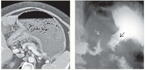 (Left) Axial CECT in a 71-year-old woman with early satiety, weight loss, and loss of appetite shows a distended stomach but a contracted antrum and thickened wall  . Infiltration of the perigastric fat, as well as enlarged lymph nodes . Infiltration of the perigastric fat, as well as enlarged lymph nodes  , indicate spread beyond the gastric wall. (Right) A film from an upper GI series in the same patient illustrates the antral constricting mass , indicate spread beyond the gastric wall. (Right) A film from an upper GI series in the same patient illustrates the antral constricting mass  and partial outlet obstruction, along with destruction of the mucosal pattern and partial outlet obstruction, along with destruction of the mucosal pattern  of the stomach. of the stomach. |
TERMINOLOGY
Definitions
Malignancy arising from gastric mucosa
IMAGING
General Features
Best diagnostic clue
Polypoid or circumferential mass with no peristalsis through lesion
Morphology
Polypoid, ulcerated, infiltrative lesions
Fluoroscopic Findings
Early (elevated, superficial, shallow)
Type 1: Elevated polypoid lesion protruding > 5 mm into lumen
Type 2: Superficial plaque-like lesion with mucosal nodularity/ulceration
Type 3: Shallow, irregular ulcer crater with adjacent nodular mucosa, clubbing/fusion/amputation of radiating folds
Advanced
Polypoid cancer can be lobulated or fungatingRelated posts:

Stay updated, free articles. Join our Telegram channel

Full access? Get Clinical Tree


