Gastric Metastases and Lymphoma
Michael P. Federle, MD, FACR
R. Brooke Jeffrey, MD
Key Facts
Imaging
Best diagnostic clue
“Bull’s-eye” lesions on imaging
Best imaging tool
Helical CT, barium (single/double) contrast studies
Top Differential Diagnoses
Gastric carcinoma
Gastric stromal tumor (leiomyosarcoma)
Gastritis (erosive type)
Pancreatitis (extrinsic inflammation)
Pathology
Classified into 2 types based on pathology
Low-grade MALT lymphoma
High-grade or advanced lymphoma
Clinical Issues
Complications
Upper GI bleeding and perforation in ulcerated lesions
Antral lesion + pyloric extension: Outlet obstruction
Treatment
Chemotherapy, surgical resection of lesions if upper GI bleed or perforation
Prognosis: Poor
Diagnostic Checklist
Check for history of primary cancer/H. pylori gastritis
Image interpretation pearls
Imaging important to suggest and stage malignancy, but biopsy often required
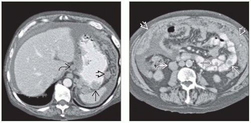 (Left) Axial CECT in a 69-year-old man shows widespread metastases from the patient’s known metastatic melanoma, including the gastric wall  , lymph nodes , lymph nodes  , and omentum , and omentum  . (Right) Axial CECT in the same patient again illustrates classic widespread metastases from melanoma, here involving the small bowel . (Right) Axial CECT in the same patient again illustrates classic widespread metastases from melanoma, here involving the small bowel  , lymph nodes , lymph nodes  , and omentum , and omentum  , with both nodular and diffuse metastases seen. In addition, the left ureter was obstructed due to a ureteral/retroperitoneal metastasis. , with both nodular and diffuse metastases seen. In addition, the left ureter was obstructed due to a ureteral/retroperitoneal metastasis. |
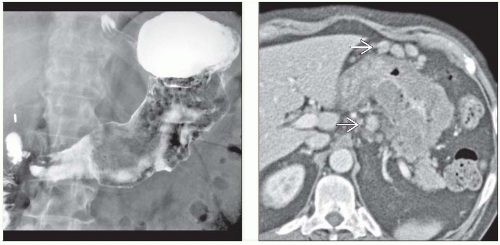 (Left) Upper GI in a 70-year-old man with weight loss and dyspepsia reveals distortion and blunting of the gastric folds. In spite of what appears to be diffuse involvement of the stomach, there is no outlet obstruction, and the stomach is distensible. (Right) Axial CECT in the same patient shows massive thickening of the gastric wall of soft tissue attenuation. Note the extensive regional lymphadenopathy and omental tumor deposits  . These findings are typical of gastric lymphoma. . These findings are typical of gastric lymphoma. |
TERMINOLOGY
Definitions
Gastric metastases from primary cancer
Lymphoma: Malignant gastric tumor of B lymphocytes
IMAGING
General Features
Best diagnostic clue
“Bull’s-eye” lesions on imaging
Fluoroscopic Findings
Fluoroscopic-guided barium study
Malignant melanoma metastases
Solitary/multiple discrete submucosal masses
“Bull’s-eye” or “target” lesions: Centrally ulcerated submucosal masses
“Spoke-wheel” pattern: Radiating superficial fissures from central ulcer
Giant cavitated lesion: Large collection of barium (5-15 cm) communicating with lumen
Small or large lobulated masses
Breast carcinoma metastases
Lobular breast cancer: Linitis plastica or “leather bottle” appearance (loss of distensibility of antrum and body + thickened irregular folds)Related posts:

Stay updated, free articles. Join our Telegram channel

Full access? Get Clinical Tree


