Gastric Ulcer
Michael P. Federle, MD, FACR
Key Facts
Imaging
Benign gastric ulcer
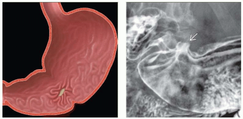
Sharply defined mucosal defect (ulcer); smooth, even, radiating folds to edge of ulcer crater
Projects beyond expected contour of stomach (UGI and CT)
Usually on lesser curve, posterior wall, or antrum
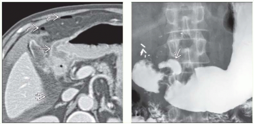
CT may show extravasation of gas and oral contrast (lesser sac or peritoneal cavity)
Malignant ulcer
Uneven shape; irregular or asymmetric edges; interruption and clubbing of radiating folds
Does not project beyond contour of stomach
CT may show mets to nodes, peritoneum, liver
Upper GI series to show ulcer
CT to show complications (± ulcer itself)
CT gastroscopy (in experienced hands) may compete with endoscopy
Sump ulcers: Distal 1/2 of greater curvature (NSAIDs)
Incisura defect: Smooth or narrow indentation on curvature opposite ulcer (muscle contraction)
Top Differential Diagnoses
Gastritis
Gastric GIST
Gastric metastases and lymphoma
Pathology
2 major risk factors: H. pylori (60-80%) and NSAIDs (20%)
Clinical Issues
Benign (95%), malignant (5%)
Often multiple: 20-30% prevalence
Hemorrhage, perforation, gastric outlet obstruction and fistula
TERMINOLOGY
Abbreviations
Peptic ulcer disease
Definitions
Inflammatory erosion of gastric mucosa
IMAGING
General Features
Best diagnostic clue
Sharply marginated barium collection with folds radiating to edge of ulcer crater on upper GI series
Location
Benign gastric ulcer
Usually lesser curvature or posterior wall of antrum or body
3-11% on greater curvature, 1-7% on anterior wall
Malignant gastric ulcer
Usually greater curvature
Morphology
Same criteria are used for findings on upper GI series, CT “virtual gastroscopy,” and endoscopy
Benign ulcer
Sharply defined mucosal defect (ulcer); smooth, even, radiating folds to edge of ulcer crater
Project beyond expected contour of stomach (UGI and CT)
Usually on lesser curve, posterior wall, or antrum
Malignant ulcer
Uneven shape; irregular or asymmetric edges; interruption and clubbing of radiating folds
Does not project beyond contour of stomach
Radiographic Findings
Upper GI series
Benign gastric ulcer, profile view
Ulcer crater: Round or ovoid collections of barium
Hampton line: Thin radiolucent line separating barium in gastric lumen from barium in crater
Ulcer mound: Smooth, bilobed hemispheric mass projecting into lumen on both sides of ulcer; outer borders form obtuse, gently-sloping angles with adjacent gastric wall (edema or inflammation)
Ulcer collar: Radiolucent rim of edematous mucosa around ulcer
Ulcer projecting beyond gastric wall
Smooth, symmetric radiating folds to edge of ulcer crater
Incisura defect: Smooth or narrow indentation on curvature opposite ulcer (muscle contraction)
Stay updated, free articles. Join our Telegram channel

Full access? Get Clinical Tree


